
Update of cochlear densitometry as a radiological diagnosis of otosclerosis
DOI:
https://doi.org/10.53903/01212095.244Keywords:
Densitometry, Otosclerosis, Multidetector computed tomography, Hearing lossAbstract
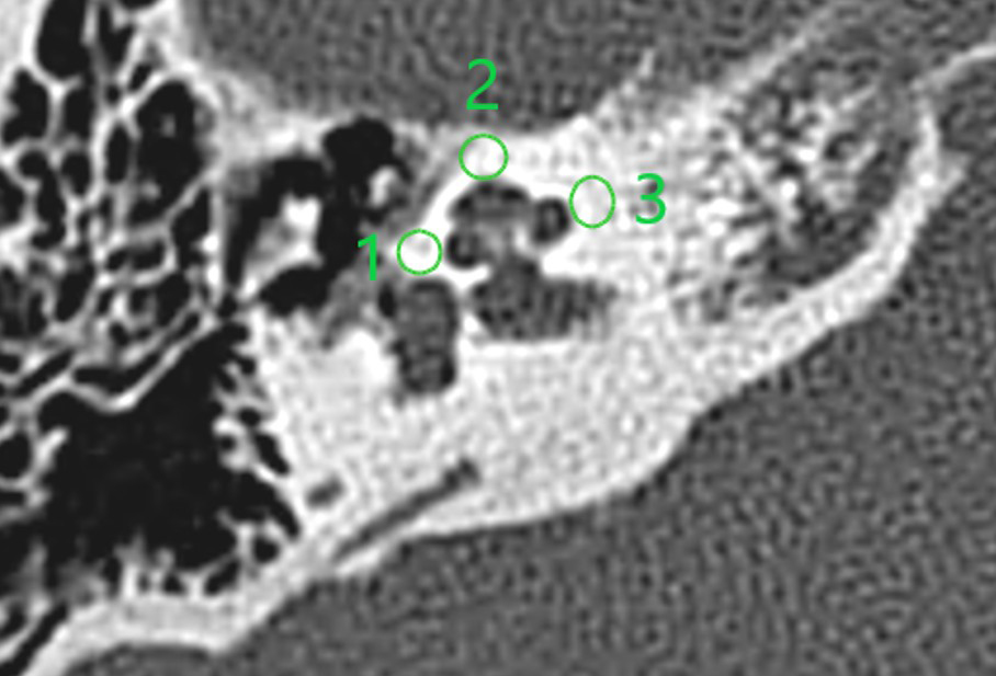
Otosclerosis is a disease of unknown etiology, frequently causing hearing loss as a result of osteodystrophy of the labyrinthine capsule that generates hearing loss due to fixation of the stapes, or to formation of foci of bone sclerosis at the endocochlear level. Diagnosis is generally clinical; however high-resolution computed tomography of the temporal bone is the imaging method of choice. The technique used is cochlear densitometry, which consists of the quantitative measurement of tissue density of specific areas in the affected osseous structures of the inner ear, by placing regions of interest (ROI).
Downloads
References
López-Galletti N, Surur A, Zernotti M, Marangoni A, Tiscornia P, Simez I. Valor de la tomografía computada multicorte en la evaluación de la otoesclerosis inicial o temprana. Revista Argentina de Diagnóstico por Imágenes. 2015;4(11):38-45
Lee TC, Aviv RI, Chen JM, Nedzelski JM, Fox AJ, Symos SP. (2009). CT grading of otosclerosis. Am J Neuroradiol. 2009;30(7):1435-9
Gredilla Molinero J, Manchego M, Nuria N, Arévalo M, Grande M. (2016). Actualización en el diagnóstico radiológico de la otosclerosis. Radiología. 2016;58(4):246-56
Zernotti ME. Contribución de la tomografía computada con densitometría ósea en el diagnóstico de la otoesclerosis [Tesis]. Universidad Católica de Córdoba; 2003
Casas JS, Rodríguez D, Miranda G, Grazia J. (2016). Otoesclerosis: revisión de aspectos etiopatogénicos, clínico-demográficos e imagenológicos. Rev. chil. radiol. [internet]. 2016;22(3):108-13. http://dx.doi.org/10.1016/j.rchira.2016.08.002
Puiggrós IV, Granell E, Calvo C, Bohé M, Orús C, Quer M. (2019). Diagnostic utility of labyrinth capsule bone density in the diagnosis of otosclerosis with high resolution tomography. Acta Otorrinolaringol Esp (Engl Ed). 2020;71(4):242-8 https://doi.org/10.1016/j.otorri.2019.09.004
Kawase S, Naganawa S, Sone M, Ikeda M, Ishigaki T. Relationship between CT densitometry with a slice thickness of 0.5 mm and audiometry in otosclerosis. Eur Radiol. 2006;16(6):1367-73. https://doi.org/10.1007/s00330-005-0128-7
Purohit B, Hermans R, Op K. (2014). Imaging in otosclerosis: A pictorial review. Insights Imaging. 2014;5(2):245-52. https://doi.org/10.1007/s13244-014-0313-9
Bozorg Grayeli A, Saint C, Imauchi Y, Cyna-Gorse F, Ferrary E, Sterkers O. Temporal bone density measurements using CT in otosclerosis. Acta Otolaryngol. 2004;124(10):1136-40. https://doi.org/10.1080/00016480410018188
Downloads
Published
How to Cite
Issue
Section
License
Copyright (c) 2025 Revista Colombiana de Radiología

This work is licensed under a Creative Commons Attribution-NonCommercial-ShareAlike 4.0 International License.
La Revista Colombiana de Radiología es de acceso abierto y todos sus artículos se encuentran libre y completamente disponibles en línea para todo público sin costo alguno.
Los derechos patrimoniales de autor de los textos y de las imágenes del artículo como han sido transferidos pertenecen a la Asociación Colombiana de Radiología (ACR). Por tanto para su reproducción es necesario solicitar permisos y se debe hacer referencia al artículo de la Revista Colombiana de Radiología en las presentaciones o artículos nuevos donde se incluyan.


