
Caudal regression syndrome type 2 associated with tethered cord: review of magnetic resonance images. A case report
DOI:
https://doi.org/10.53903/01212095.336Keywords:
Caudal regression syndrome, Neural tube defects, Sacral agenesis, Diabetes mellitusAbstract
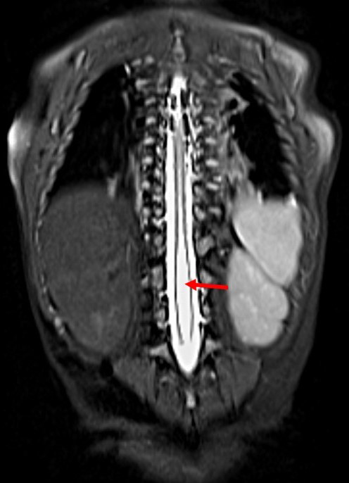
Caudal regression syndrome, also known as caudal dysplasia sequence, is a rare severe congenital anomaly characterized by variable extension agenesis of the distal vertebral bodies (sacrococcygeal or lumbosacrococcygeal), which is frequently associated with other musculoskeletal malformations of the pelvis and lower extremities; neurological, orthopedic, gastrointestinal, genitourinary and cardiac abnormalities have also been described. In the evaluation of caudal regression syndrome, magnetic resonance imaging MRI) allows an adequate diagnosis not only of congenital anomalies of the spinal cord and spine, but also of most associated anomalies in the pelvic region. We present the case of a 1-year-old male with MRI of caudal regression syndrome type 2, tethered spinal cord, intradural lipoma, presence of butterfly vertebrae, hemivertebrae and duplication. This article describes the imaging findings of caudal regression syndrome as well as the differences in its classification.
Downloads
References
Luque H, Fernández R, Tuca M, Luco M, De Barbieri F, Tapia J. Síndrome de regresión caudal. Caso clínico. Rev chil pediatr. 2010;81(2). https://doi.org/10.4067/s0370-41062010000200007
Zepeda TJ, García MM, Morales SJ, Pantoja HM, Espinoza GA. Secuencia de regresión caudal: caso clínico-radiológico. Rev chil pediatr. 2015;86(6):430-5. https://doi.org/10.1016/j.rchipe.2015.07.021
Trapp B, Freddi TAL, Hans MOM, Calixto LFT, Fujino E, Rojas LCA, et al. A practical approach to diagnosis of spinal dysraphism. Radiographics. 2021;41(2):559-75. https://doi.org/10.1148/rg.2021200103
Krishnan V, Jaganathan S, Jayappa S, et al. Clinical and radiological evaluation of caudal regression syndrome. Pediatr Radiol. 2024;54(9):1451-61. https://doi.org/10.1007/s00247-024-05945-1
Rojansky N, Fasouliotis SJ, Ariel I, Nadjari M. Extreme caudal agenesis. Possible drug-related etiology? J Reprod Med. 2002;47(3):241-5.
Nievelstein RA, Valk J, Smit LM, Vermeij-Keers C. MR of the caudal regression syndrome: embryologic implications. Am J Neuroradiol. 1994;15(6):1021-9.
Singh SK, Singh RD, Sharma A. Caudal regression síndrome, case report and review of literature. Ped Surgery Int. 2005;21:578-81. https://doi.org/10.1007/s00383-005-1451-4
Unsinn KM, Geley T, Freund MC, Gassner I. US of the spinal cord in newborns: Spectrum of normal findings, variants, congenital anomalies, and acquired diseases. Radio-Graphics. 2000;20(4):923-38. https://doi.org/10.1148/radiographics.20.4.g00jl06923
Boruah DK, Dhingani D, Achar S, Prakash A, Augustine A, Sanyal S, et al. Magnetic resonance imaging analysis of caudal regression syndrome and concomitant anomalies in pediatric patients. J Clin Imaging Sci. 2016;6:36. https://doi.org/10.4103/2156-7514.190892
Diel J, Ortiz O, Losada RA, Price DB, Hayt MW, Katz DS. The sacrum: pathologic spectrum, multimodality imaging, and subspecialty approach. Radiographics. 2001;21(1):83-104. https://doi.org/|10.1148/radiographics.21.1.g01ja0883
Baxi L, Warren W, Collins MH, Timor-Tritsch IE. Early detection of caudal regression syndrome with transvaginal scanning. Obstet Gynecol. 1990;75(3 Pt 2):486-9.
Downloads
Published
How to Cite
Issue
Section
License
Copyright (c) 2025 Revista Colombiana de Radiología

This work is licensed under a Creative Commons Attribution-NonCommercial-ShareAlike 4.0 International License.
La Revista Colombiana de Radiología es de acceso abierto y todos sus artículos se encuentran libre y completamente disponibles en línea para todo público sin costo alguno.
Los derechos patrimoniales de autor de los textos y de las imágenes del artículo como han sido transferidos pertenecen a la Asociación Colombiana de Radiología (ACR). Por tanto para su reproducción es necesario solicitar permisos y se debe hacer referencia al artículo de la Revista Colombiana de Radiología en las presentaciones o artículos nuevos donde se incluyan.


