
Association between chest ct findings and prognosis in patients with SARS-CoV-2 pneumonia over a 4-week follow-up
DOI:
https://doi.org/10.53903/01212095.339Keywords:
SARS-CoV-2, Tomography, X-Ray Computed, PrognosisAbstract
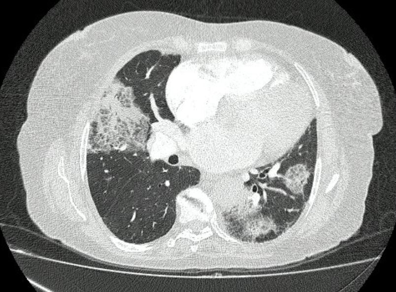
Introduction: The SARS-CoV-2 pandemic has caused millions of deaths, with SARS-CoV-2 pneumonia being a leading cause of acute respiratory infection. Our objective is to evaluate the association between chest CT findings and prognosis in patients hospitalized for this infection. Methods: We included 360 adult patients from a fourth level center, diagnosed with COVID-19 by RT-PCR, who had a chest CT scan at the beginning of their hospitalization. We assessed by means of logistic regression the relationship between imaging findings and survival. Results: The most frequent findings were ground glass pattern (87%), followed by consolidation (27%), cobblestone pattern (18%) and organizing pneumonia (6%). Most patients survived (77.5%), while 16.9% died within four weeks. The presence of alterations in the diagnostic imaging represented a tendency to be risk factors for death, finding that consolidation was associated with an increased risk of mortality by 10%. Discussion: The detection of alterations or semiological findings on chest CT is associated with worse prognosis within the first 4 weeks for SARS-CoV-2 infected patients. The results highlight that early identification of certain imaging findings could improve severity prediction and guide clinical decisions. Going forward, it is recommended to evaluate the chronological evolution of findings to optimize the management of patients with SARS-CoV-2 infection.
Downloads
References
Weekly operational update on COVID-19 [nota de prensa]. 2021, 4 de septiembre. Disponible en: https://www.who.int/publications/m/item/weekly-operational-updateon-covid-19-4-august-2021.
Botero-Rodríguez F, Franco O, Gómez C. Pandemic’s glossary: The ABC of coronavirus concepts [Glosario para una pandemia: el ABC de los conceptos sobre el coronavirus]. Biomédica. 2020;40(Supl. 2):16-26. https://doi.org/10.7705/biomedica.5605
Rodríguez-Morales AJ, Cardona-Ospina JA, Gutiérrez- Ocampo E, Villamizar-Pena R, Holguín-Rivera Y, Escalera-Antezana JP, et al. Clinical, laboratory and imaging features of COVID-19: A systematic review and meta-analysis. Travel Med Infect Dis. 2020 Mar;(March):101623. https://doi.org/10.1016/j.tmaid.2020.101623
Zhao W, Zhong Z, Xie X, Yu Q, Liu J. Relation between chest CT findings and clinical conditions of coronavirus disease (COVID-19) pneumonia: A multicenter study. Am J Roentgenol. 2020:214(5):1072-7. https://doi.org/10.2214/AJR.20.22976
Bernheim A, Mei X, Huang M, Yang Y, Fayad ZA, Zhang N, et al. Chest CT Findings in coronavirus disease-19 (COVID-19): Relationship to duration of infection. Radiology. 2020;295(3):200463. https://doi.org/10.1148/radiol.2020200463
Pan F, Ye T, Sun P, Gui S, Liang B, Li L, et al. Time course of lung changes on chest CT during recovery from 2019 novel coronavirus (COVID-19) pneumonia. Radiology. 2020;295(3):715-21. https://doi.org/10.1148/radiol.2020200370
Kwee TC, Kwee RM. Chest CT in COVID-19: What the Radiologist needs to know. RadioGraphics. 2020;40(7):1848-65. https://doi.org/10.1148/rg.2020200159
Abbasian Ardakani A, Acharya UR, Habibollahi S, Mohammadi A. COVIDiag: a clinical CAD system to diagnose COVID-19 pneumonia based on CT findings. Eur. Radiol. 2020;31(1):121-30. https://doi.org/10.1007/s00330-020-07087-y
Lyu P, Liu X, Zhang R, et al. The performance of chest CT in evaluating the clinical severity of COVID-19 pneumonia: identifying critical cases based on CT characteristics. Invest Radiol. 2020;55(7):412-421. https://doi.org/10.1097/RLI.0000000000000689
Li K, Fang Y, Li W, et al. CT image visual quantitative evaluation and clinical classification of coronavirus disease (COVID-19). Eur Radiol. 2020;30(8):4407-16. https://doi.org/10.1007/s00330-020-06817-6
Gong K, Wu D, Arru CD, Homayounieh F, Neumark N, Guan J, et al. A multi-center study of COVID-19 PATIENT prognosis using DEEP learning-based CT image analysis and electronic health records. Eur J Radiol. 2021;139:109583. https://doi.org/10.1016/j.ejrad.2021.109583
Wang Y, Dong C, Hu Y, Li C, Ren Q, Zhang X, et al. Temporal changes of CT findings in 90 patients with COVID-19 pneumonia: A longitudinal study. Radiology. 2020;296(2):E55-E64. https://doi.org/10.1148/radiol.2020200843
Hansell DM, Bankier AA, MacMahon H, McLoud TC, Müller NL, Remy J. Fleischner Society: glossary of terms for thoracic imaging. Radiology. 2008;246(3):697-722. https://doi.org/10.1148/radiol.2462070712
Mehri A, Sotoodeh Ghorbani S, Farhadi-Babadi K, Rahimi E, Barati Z, Taherpour N, et al. Risk factors associated with severity and death from COVID-19 in Iran: A systematic review and meta-analysis study. J Intensive Care Med. 2023;38(9):825-37. https://doi.org/10.1177/08850666231166344
Anderegg N, Panczak R, Egger M, et al. Survival among people hospitalized with COVID-19 in Switzerland: a nationwide population-based analysis. BMC Med. 2022;20(1):164. https://doi.org/10.1186/s12916-022-02364-7
Yuan M, Yin W, Tao Z, et al. Association of radiologic findings with mortality of patients infected with 2019 novel coronavirus in Wuhan, China. PLoS One. 2020;15(3):e0230548. https://doi.org/10.1371/journal.pone.0230548
Colombi D, Bodini FC, Petrini M, et al. Well-aerated lung on admitting chest CT to predict adverse outcome in COVID-19 pneumonia. Radiology. 2020;296(2):E86-E96. https://doi.org/10.1148/radiol.2020201433
Downloads
Published
How to Cite
Issue
Section
License
Copyright (c) 2025 Revista Colombiana de Radiología

This work is licensed under a Creative Commons Attribution-NonCommercial-ShareAlike 4.0 International License.
La Revista Colombiana de Radiología es de acceso abierto y todos sus artículos se encuentran libre y completamente disponibles en línea para todo público sin costo alguno.
Los derechos patrimoniales de autor de los textos y de las imágenes del artículo como han sido transferidos pertenecen a la Asociación Colombiana de Radiología (ACR). Por tanto para su reproducción es necesario solicitar permisos y se debe hacer referencia al artículo de la Revista Colombiana de Radiología en las presentaciones o artículos nuevos donde se incluyan.


