
First trimester diagnosis of fetal gastroschisis, case report and literature revisión
DOI:
https://doi.org/10.53903/01212095.382Keywords:
Gastroschisis, Ultrasonography, Abdominal wall, Prenatal diagnosisAbstract
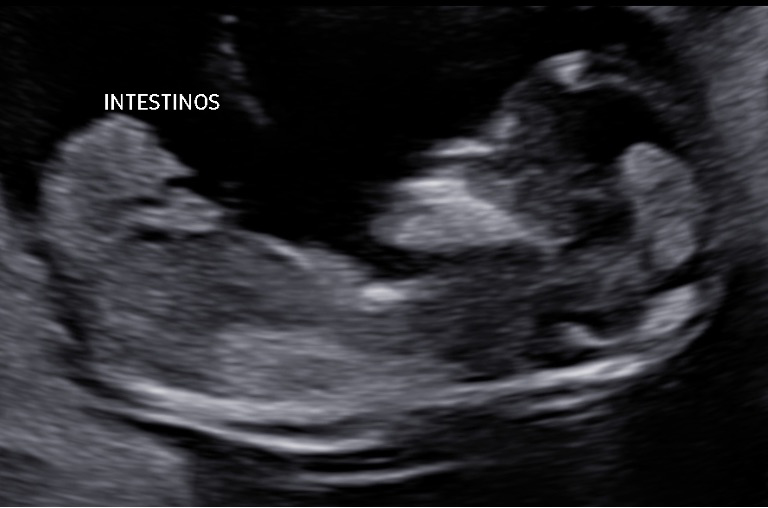
The spectrum of fetal abdominal wall defects includes omphalocele, gastroschisis, pentalogy of Cantrell, bladder and cloacal exstrophy, among others. These pathologies are usually diagnosed during second-trimester ultrasounds, generally during the anatomical detail sonogram performed
between 20 and 24 weeks. However, careful assessment by the sonographer, starting in the embryonic phase, allows for the detection of these anomalies in the first trimester. It is important to differentiate the main morphological characteristics of these wall defects, since the suggestion of any of them as a diagnosis entails a chain of decision-making, involving the medical team and the expectant parents. Below, a case of gastroschisis in a primiparous woman who came for a first-trimester screening ultrasound is presented. The imaging findings were unequivocal and typical of the condition, which allowed for an early diagnosis and the decision-making considered pertinent by the parents.
Downloads
References
Revels J, Wang S, Nasrullah A, Revzin M, Iyer R, Deutsch G, et al. An algorithmic approach to complex fetal abdominal wall defects. Am J Roentgenol. 2020;214(1):218- 31. https://doi.org/10.2214/AJR.19.21627.
Arif RH, Mahmoud LB, Ali ABE, et al. Gastroschisis: anatomic defects, etiopathogenesis, treatment, and prognosis. Newborn. 2022;1(3):287-96. https://doi.org/10.5005/jp-journals-11002-0041.
Torfs CP, Ellie E, Oecchsli T, et al. A population-based study of gastros- chisis: demographic, pregnancy, and lifestyle risk factors. Teratology. 1994;50:44-53. https://doi.org/10.1002/tera.1420500107.
Syngelaki A, Chelemen T, Dagklis T, Allan L, Nicolaides KH. Challenges in the diagnosis of fetal non-chromosomal abnormalities at 11-13 weeks. Prenat Diagn. 2011;31:90-102. https://doi.org/10.1002/pd.2642.
Marquart JP, Nie Q, Gonzalez T, Jelin A, Broeckel U, Wagner A, et al. Genetics and genomics of gastroschisis, elucidating a potential genetic etiology for the most common abdominal defect: a systematic review. J. Dev. Biol. 2024;12(4):34. https://doi.org/10.3390/jdb12040034.
Al-Kaff A, MacDonald S, Kent N, Burrows J, Skarsgard E, Hutcheon J, et al. Delivery planning for pregnancies with gastroschisis: Findings from a prospective national registry. Am J Obstet Gynecol. 2015;213(4):557:e1–e8. https://doi.org/10.1016/j.ajog.2015.06.048.
Palatnik A, Loichinger M, Wagner M, Peterson E. The association between gestational age at delivery, closure type and perinatal outcomes in neonates with isolated gastroschisis. J Matern Fetal Neonatal Med. 2020;33(8):1393-9. https://doi.org/10.1080/14767058.2018.1519538.
Fraga MV, Laje P, Peranteau W, Hedrick H, Khalec N, Gebb J, et al. The influence of gestational age, mode of delivery and abdominal wall closure method on the surgical outcome of neonates with uncomplicated gastroschisis. Pediatr Surg Int. 2018;34(4):415-9. https://doi.org/10.1007/s00383-018-4233-5.
Banyard D, Ramones T, Phillips S, Leys C, Rauth T, Yang E, et al. Method to our madness: An 18-year retrospective analysis on gastroschisis closure. J Pediatr Surg. 2010;45(3):579-84. https://doi.org/10.1016/j.jpedsurg.2009.08.004.
Kunz SN, Tieder S, Whitlock K, Jackson C, Avansino J. Primary fascial closure versus staged closure with silo in patients with gastroschisis: A meta-analysis. J Pediatr Surg. 2013;48(4):845-57. https://doi.org/10.1016/j.jpedsurg.2013.01.020.
Bergholz R, Boettcher M, Reinshagen K, Wenke K. Complex gastroschisis is a different entity to simple gastroschisis affecting morbidity and mortality: A systematic review and meta-analysis. J Pediatr Surg. 2014;49(10):1527-32. https://doi.org/10.1016/j.jpedsurg.2014.08.001.
Zalles-Vidal C, Vega M, Valadez M, Cabrera M. Late prematurity with gastroschisis and severe hypoalbuminemia. Bol Med Hosp Infant Mex. 2015;72(5):339-45. https://doi.org/10.1016/j.bmhimx.2015.08.004.
Downloads
Published
How to Cite
Issue
Section
License
Copyright (c) 2025 Revista Colombiana de Radiología

This work is licensed under a Creative Commons Attribution-NonCommercial-ShareAlike 4.0 International License.
La Revista Colombiana de Radiología es de acceso abierto y todos sus artículos se encuentran libre y completamente disponibles en línea para todo público sin costo alguno.
Los derechos patrimoniales de autor de los textos y de las imágenes del artículo como han sido transferidos pertenecen a la Asociación Colombiana de Radiología (ACR). Por tanto para su reproducción es necesario solicitar permisos y se debe hacer referencia al artículo de la Revista Colombiana de Radiología en las presentaciones o artículos nuevos donde se incluyan.


