
Manifestaciones radiológicas de malformaciones pulmonares congénitas. Experiencia de tres hospitales en Bogotá
DOI:
https://doi.org/10.53903/01212095.67Palabras clave:
Anomalías congénitas, Tomografía computarizada multidetector, Enfermedades pulmonares, NiñoResumen
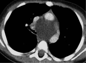
Objetivo: Describir las características radiológicas de las malformaciones congénitas pulmonares y de la vía aérea que se encuentran frecuentemente en pacientes pediátricos, de acuerdo con la experiencia de tres hospitales de Bogotá, entre 2010 y 2016. Materiales y métodos: Estudio retrospectivo, observacional y descriptivo con muestra de 27 pacientes de 5 meses de edad promedio, que cumplieron criterios de inclusión: pacientes entre los 0 meses y 17 años de edad, pacientes con diagnóstico confirmado de malformación congénita del pulmón, pacientes intervenidos quirúrgicamente por lesión pulmonar o de la vía aérea y cuyo estudio histopatológico fue compatible con malformación congénita del pulmón. Resultados: La prevalencia de las malformaciones congénitas es mayor en el sexo femenino, 80 % de los casos contaron con diagnóstico prenatal, la malformación quística adenomatoidea es la más frecuente y la principal característica radiológica es el quiste. Conclusión: La tomografía computarizada permite estudios detallados de estas malformaciones, con una mayor precisión en comparación con las técnicas convencionales como radiografía de tórax y ultrasonografía.
Descargas
Referencias bibliográficas
Biyyam DR, Chapman T, Ferguson MR, Deutsch G, Dighe MK. Congenital lung abnormalities: Embryologic features, prenatal diagnosis, and postnatal radiologicpathologic correlation. RadioGraphics. 2010;30(6):1721-38.
Lee EY, Dorkin H, Vargas SO. Congenital pulmonary malformations in pediatric patients: review and update on etiology, classification, and imaging findings. Radiol Clin North Am. 2011;49(5):921-48.
Lee EY, Boiselle PM, Cleveland RH. Multidetector CT evaluation of congenital lung anomalies 1. Radiology. 2008;247(3):632-48.
Antón-Martín P, Cuesta-Rubio MT, López-González MF, Ortiz-Movilla R, Lorente- Jareño ML, López-Rodríguez E, et al. Malformación adenomatoidea quística congénita. Rev chil. pediatría. 2011;82:129-36.
Ch’In KY, Tang MY. Congenital adenomatoid malformation of one lobe of a lung with general anasarca. Arch Pathol (Chic). 1949;48(3):221-9.
Stocker JT, Madewell JE, Drake RM. Congenital cystic adenomatoid malformation of the lung. Classification and morphologic spectrum. Hum Pathol. 1977;8(2):155-71.
Stocker JT. Congenital pulmonary airway malformation: A new name and an expanded classification of congenital cystic adenomatoid malformation of the lung. Histopathology. 2002;41(Suppl 2):424-31.
Stocker JT. Cystic lung disease in infants and children. Fetal Pediatr Pathol. 2009;28(4):155-84.
Mehta AA, Viswanathan N, Vasudevan AK, Paulose R, Abraham M. Congenital cystic adenomatoid malformation: A tertiary care hospital experience. J Clin Diagn Res. 2016;10(11):SC01-SC4.
Trotman-Dickenson B. Congenital lung disease in the adult: guide to the evaluation and management. J Thorac Imaging. 2015;30(1):46-59.
Kao SW, Zuppan CW, Young LW. AIRP Best cases in radiologic-pathologic correlation: Type 2 congenital cystic adenomatoid malformation (Type 2 congenital pulmonary airway malformation). RadioGraphics. 2011;31(3):743-8.
Williams HJ, Johnson KJ. Imaging of congenital cystic lung lesions. Paediatr Respir Rev. 2002;3(2):120-7.
Tashtoush B, Memarpour R, González J, Gleason JB, Hadeh A. Pulmonary sequestration: A 29 patient case series and review. J Clin Diagn Res. 2015;9(12):Ac05-8.
Pryce DM. Lower accessory pulmonary artery with intralobar sequestration of lung; a report of seven cases. J Pathol Bacteriol. 1946;58(3):457-67.
Clements BS, Warner JO. Pulmonary sequestration and related congenital bronchopulmonary- vascular malformations: nomenclature and classification based on anatomical and embryological considerations. Thorax. 1987;42(6):401-8.
Wei Y, Li F. Pulmonary sequestration: a retrospective analysis of 2625 cases in China. Eur J Cardiothorac Surg. 2011;40(1):e39-42.
Qian X, Sun Y, Liu D, Wu X, Wang Z, Tang Y. Pulmonary sequestration: a case report and literature review. Int J Clin Exp Med. 2015;8(11):21822-5.
Berrocal T, Madrid C, Novo S, Gutiérrez J, Arjonilla A, Gómez-León N. Congenital anomalies of the tracheobronchial tree, lung, and mediastinum: Embryology, radiology, and pathology. RadioGraphics. 2004;24(1):e17-e. Pubmed.
Heithoff KB, Sane SM, Williams HJ, Jarvis CJ, Carter J, Kane P, et al. Bronchopulmonary foregut malformations. A unifying etiological concept. AJR Am J Roentgenol. 1976;126(1):46-55.
Jeung MY, Gasser B, Gangi A, Bogorin A, Charneau D, Wihlm JM, et al. Imaging of cystic masses of the mediastinum. Radiographics. 2002;22(Spec No):S79-93.
Kaistha A, Levine J. An unusual cause of pediatric dysphagia: Bronchogenic cyst. Glob Pediatr Health. 2017;4:2333794x16686492.
Rogers LF, Osmer JC. Bronchogenic cyst. A review of 46 cases. Am J Roentgenol Radium Ther Nucl Med. 1964;91:273-90.
Abdellah O, Mohamed H, Youssef B, Abdelhak B. A case of congenital lobar emphysema in the middle lobe. J Clin Neonatol. 2013;2(3):135-7.
Latif I, Shamim S, Ali S. Congenital lobar emphysema. J Pak Med Assoc. 2016;66(2):210-2.
Jacob M, Ramesh GS, Narmadha Lakshmi K. Anesthetic management of congenital lobar emphysema in a neonate. Med J Armed Forces India. 2015;71(Suppl 1):S287-9.
Zylak CJ, Eyler WR, Spizarny DL, Stone CH. Developmental lung anomalies in the adult: Radiologic-pathologic correlation. RadioGraphics. 2002;22(suppl_1):S25-S43.
Cataneo DC, Rodrigues OR, Hasimoto EN, Schmidt Jr AF, Cataneo AJ. Congenital lobar emphysema: 30-year case series in two university hospitals. J Bras Pneumol. 2013;39(4):418-26.
Daltro P, Fricke BL, Kuroki I, Domingues R, Donnelly LF. CT of congenital lung lesions in pediatric patients. AJR Am J Roentgenol. 2004;183(5):1497-506.
Manivel JC, Priest JR, Watterson J, Steiner M, Woods WG, Wick MR, et al. Pleuropulmonary blastoma. The so-called pulmonary blastoma of childhood. Cancer. 1988;62(8):1516-26. Pubmed.
Priest JR, McDermott MB, Bhatia S, Watterson J, Manivel JC, Dehner LP. Pleuropulmonary blastoma: a clinicopathologic study of 50 cases. Cancer. 1997;80(1):147-61.
Barnard WG. Embryoma of lungs. Thorax. 1952;7(4):299-301.
Dehner LP. Pleuropulmonary blastoma is the pulmonary blastoma of childhood. Semin Diagn Pathol. 1994;11(2):144-51.
Miniati DN, Chintagumpala M, Langston C, Dishop MK, Olutoye OO, Nuchtern JG, et al. Prenatal presentation and outcome of children with pleuropulmonary blastoma. J Pediatr Surg. 2006;41(1):66-71.
Lee HJ, Goo JM, Kim KW, Im JG, Kim JH. Pulmonary blastoma: radiologic findings in five patients. Clin Imaging. 2004;28(2):113-8.
Herer B, Jaubert F, Delaisements C, Huchon G, Chretien J. Scimitar sign with normal pulmonary venous drainage and anomalous inferior vena cava. Thorax. 1988;43(8):651-2.
Chowdhury MM, Chakraborty S. Imaging of congenital lung malformations. Semin Pediatr Surg. 2015;24(4):168-75.
Keslar P, Newman B, Oh KS. Radiographic manifestations of anomalies of the lung. Radiol Clin North Am. 1991;29(2):255-70.
Panicek DM, Heitzman ER, Randall PA, Groskin SA, Chew FS, Lane EJ, Jr., et al. The continuum of pulmonary developmental anomalies. Radiographics. 1987;7(4):747-72.
Descargas
Publicado
Cómo citar
Número
Sección
Licencia

Esta obra está bajo una licencia internacional Creative Commons Atribución-NoComercial-CompartirIgual 4.0.
La Revista Colombiana de Radiología es de acceso abierto y todos sus artículos se encuentran libre y completamente disponibles en línea para todo público sin costo alguno.
Los derechos patrimoniales de autor de los textos y de las imágenes del artículo como han sido transferidos pertenecen a la Asociación Colombiana de Radiología (ACR). Por tanto para su reproducción es necesario solicitar permisos y se debe hacer referencia al artículo de la Revista Colombiana de Radiología en las presentaciones o artículos nuevos donde se incluyan.


