
Leiomyosarcoma of the inferior vena cava. What to look for in the images?. Presentation of a case
DOI:
https://doi.org/10.53903/01212095.162Keywords:
Leiomyosarcoma, Inferior vena cava, Multidetector computed tomographyAbstract
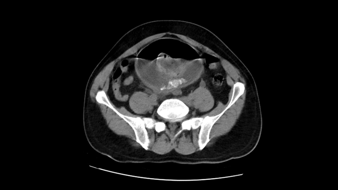
Inferior vena cava leiomyosarcoma (IVC) is a low-growing, malignant mesenchymal tumor that arises in the smooth muscle cells of the tunica media of the vascular wall and has a generally por prognosis. Images play a crucial role in the diagnostic approach and in surgical planning, therefore, recognizing the most frequent findings is necessary in radiological practice. We describe a case that outlines the most typical findings in different diagnostic modalities including tomography, magnetic resonance imaging and PET-CT, with the aim of recognizing the characteristics that can lead to an earlier diagnosis and therefore to favor patient survival.
Downloads
References
Mastoraki A, Leotsakos G, Mastoraki S, Papanikolaou IS, Danias N, Smyrniotis V, et al. Challenging diagnostic and therapeutic modalities for leiomyosarcoma of inferior vena cava. Int J Surg Lond Engl. 2015;13:92-5. doi: 10.1016/j.ijsu.2014.11.051
Bednarova I, Frellesen C, Roman A, Vogl TJ. Case 257: Leiomyosarcoma of the inferior vena cava. Radiology. 2018;288(3):901-8. doi: 10.1148/radiol.2018160821
Alkhalili E, Greenbaum A, Langsfeld M, Marek J, Rana MA, Glew R, et al. Leiomyosarcoma of the inferior vena cava: A case series and review of the literature. Ann Vasc Surg. 2016;33:245-51. doi: 10.1016/j.avsg.2015.10.016
Monteagudo Cortecero J, Guirau Rubio MD, Payá Romá A. Leiomyosarcoma of the inferior vena cava: AIRP best cases in radiologic-pathologic correlation. RadioGraphics. 2015;35(2):616-20. doi: 2020-10-08 21:37:22
Bonura A, Saade C, Sharma P. Leiomyosarcoma of the inferior vena cava. Australas Radiol. 2006;50(4):395-9. doi: 10.1111/j.1440-1673.2006.01611.x
Gage MJ, Patel AV, Koenig KL, Newman E. Non-vena cava venous leiomyosarcomas: a review of the literature. Ann Surg Oncol. 2012;19(11):3368-74. doi: 10.1245/s10434- 012-2379-2
Sulpice L, Rayar M, Levi Sandri G-B, de Wailly P, Henno S, Turner K, et al. Leiomyosarcoma of the inferior vena cava. J Visc Surg. 2016;153(3):161-5. doi: 10.1016/j.jviscsurg.2015.11.002
Xu J, Velayati A, Berger BJ, Liu M, Cheedella NKS, Gotlieb V. Leiomyosarcoma of the inferior vena cava in an HIV-positive adult patient: A case report and review of the literature. Am J Case Rep. 2017;18:1160-5. doi: 10.12659/ajcr.905787
Ceyhan M, Danaci M, Elmali M, Ozmen Z. Leiomyosarcoma of the inferior vena cava. Diagn Interv Radiol Ank Turk. 2007;13(3):140-3.
Singh N, Shivdasani D, Karangutkar S. Rare case of primary inferior vena cava leiomyosarcoma on F-18 fluorodeoxyglucose positron emission tomography-computed tomography scan: Differentiation from nontumor thrombus in a background of procoagulant state. Indian J Nucl Med IJNM Off J Soc Nucl Med India. 2014;29(4):246-8. doi: 10.4103/0972-3919.142629
Webb EM, Wang ZJ, Westphalen AC, Nakakura EK, Coakley FV, Yeh BM. Can CT features differentiate between inferior vena cava leiomyosarcomas and primary retroperitoneal masses? Am J Roentgenol. 2013;200(1):205-9. doi: 10.2214/ AJR.11.7476
Ganeshalingam S, Rajeswaran G, Jones RL, Thway K, Moskovic E. Leiomyosarcomas of the inferior vena cava: diagnostic features on cross-sectional imaging. Clin Radiol. 2011;66(1):50-6. doi: 10.1016/j.crad.2010.08.004
Zhou X, Wang M, Li S, Cai H, Liang L, Li Z-P, et al. A case of a huge inferior vena cava leiomyosarcoma: Precise preoperative evaluation with gadobutrol-enhanced MRI. Cancer Manag Res. 2020;12:7929-39. doi: 10.2147/CMAR.S258990
Mu D, Wang D, Zhou K, Zhu B. Radiographic features of intraluminal leiomyosarcoma of the inferior vena cava: an atypical case report. Abdom Imaging. 2011;36(5):586-9. doi: 10.1007/s00261-010-9673-x
Hemant D, Krantikumar R, Amita J, Chawla A, Ranjeet N. Primary leiomyosarcoma of inferior vena cava, a rare entity: Imaging features. Australas Radiol. 2001;45(4):448- 51. doi: 10.1046/j.1440-1673.2001.00955.x
Engelbrecht M, Akin O, Dixit D, Schwartz L. Bland and tumor thrombi in abdominal malignancies: magnetic resonance imaging assessment in a large oncologic patient population. Abdom Imaging. 2011;36(1):62-8. doi: 10.1007/s00261-010-9608-6
Smillie RP, Shetty M, Boyer AC, Madrazo B, Jafri SZ. Imaging evaluation of the inferior vena cava. RadioGraphics. 2015;35(2):578-92. doi: 10.1148/rg.352140136
Ito H, Hornick JL, Bertagnolli MM, George S, Morgan JA, Baldini EH, et al. Leiomyosarcoma of the inferior vena cava: survival after aggressive management. Ann Surg Oncol. 2007;14(12):3534-41. doi: 10.1245/s10434-007-9552-z
Sephien A, Mousa MS, Bui MM, Kedar R, Thomas K. Leiomyosarcoma of the inferior vena cava with hepatic and pulmonary metastases: Case report. J Radiol Case Rep. 2019;13(5):30-40. doi: 10.3941/jrcr.v13i5.3641
Teixeira FJR, do Couto Netto SD, Perina AL de F, Torricelli FCM, Ragazzo Teixeira L, Zerati AE, et al. Leiomyosarcoma of the inferior vena cava: Survival rate following radical resection. Oncol Lett. 2017;14(4):3909-16. doi: 10.1245/s10434-007-9552-z
Jeong S, Han Y, Cho Y-P, Kwon T-W. Clinical outcomes of surgical resection for leiomyosarcoma of the inferior vena cava. Ann Vasc Surg. 2019;61:377-83. doi: 10.1016/j.avsg.2019.05.053
Wachtel H, Gupta M, Bartlett EK, Jackson BM, Kelz RR, Karakousis GC, et al. Outcomes after resection of leiomyosarcomas of the inferior vena cava: a pooled data analysis of 377 cases. Surg Oncol. 2015;24(1):21-7. doi: 10.1016/j.suronc.2014.10.007
Downloads
Published
How to Cite
Issue
Section
License

This work is licensed under a Creative Commons Attribution-NonCommercial-ShareAlike 4.0 International License.
La Revista Colombiana de Radiología es de acceso abierto y todos sus artículos se encuentran libre y completamente disponibles en línea para todo público sin costo alguno.
Los derechos patrimoniales de autor de los textos y de las imágenes del artículo como han sido transferidos pertenecen a la Asociación Colombiana de Radiología (ACR). Por tanto para su reproducción es necesario solicitar permisos y se debe hacer referencia al artículo de la Revista Colombiana de Radiología en las presentaciones o artículos nuevos donde se incluyan.


