
Atypical giant hepatic hemangioma. A case report
DOI:
https://doi.org/10.53903/01212095.185Keywords:
Hemangioma, Tomography Interventional radiology, LiverAbstract
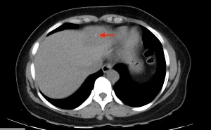
Giant hepatic hemangiomas are lesions included in the atypical hemangiomas category. In most cases they have a benign behavior, predominantly present in women with ages ranging from 20 to 60, and are commonly located in the right liver lobe with a size larger than 4 cm. The diagnosis is usually made incidentally because they generally don´t produce any symptoms; however, when they are large they tend to manifest persistent abdominal symptoms. In images, among the most common findings in ultrasonography are: hyperechoic lesions with peripheral feeding vessels and posterior acoustic reinforcement; on CT the most frequent findings are heterogeneous lesions with a markedly hypoattenuating central area that has nodular peripheral enhancement in the arterial phase after intravenous contrast administration; some atypical findings include hemorrhage, thrombus, calcifications, capsules or pseudocapsules. Although most of these lesions only require observation, some patients require treatment, either surgical (enucleation, resection) or interventional radiology procedures such as angiographic selective embolization.
Downloads
References
- Vilgrain, V., Boulos, L., Vullierme, M., Denys, A., Terris, B. and Menú, Y., 2021. Imaging of Atypical Hemangiomas of the Liver with Pathologic Correlation.
-Moctezuma-Velázquez, C., López-Arce, G., Martínez-Rodríguez, L., Escalona-Huerta, C., Chapa-Ibargüengoitia, M. and Torre, A., 2022. Hemangioma hepático gigante versus hemangioma hepático convencional: características clínicas, factores de riesgo y manejo.
- Coumbaras M, Wendum D, Monnier-Cholley L, Dahan H.,2002, CT and MR Imaging Features of Pathologically Proven Atypical Giant Hemangiomas of the Liver: American Journal of Roentgenology: Vol. 179, No. 6 (AJR)
Bajenaru, N., Balaban, V., Săvulescu, F., Campeanu, I., & Patrascu, T. (2015). Hepatic hemangioma -review-. Journal of medicine and life, 8 Spec Issue (Spec Issue), 4–11.
- Jang, H., Kim, T., Lim, H., Park, S., Sim, J., Kim, H. and Lee, J., 2003. Hepatic Hemangioma: Atypical Appearances on CT, MR Imaging, and Sonography. American Journal of Roentgenology, 180(1), pp.135-141.
- Díaz Rubia L, García Verdejo FJ. Hemangioma hepático gigante tratado con embolización. RAPD Online. 2019;42(4):148-50
Alberto Castrillón G, del Pilar Montoya M, Andrés Soto J. Giant hepatic cavernous hemangiomas: spiral computed tomography findings in 21 patients. original articles. Rev Colomb Radiol. 2010;21(2):1–5.
Vassiou, K., Rountas, H., Liakou, P. et al. Embolization of a Giant Hepatic Hemangioma Prior To Urgent Liver Resection. Case Report and Review of the Literature. Cardiovasc Intervent Radiol 30, 800–802 (2007).
Downloads
Published
How to Cite
Issue
Section
License
Copyright (c) 2023 Revista Colombiana de Radiología

This work is licensed under a Creative Commons Attribution-NonCommercial-ShareAlike 4.0 International License.
La Revista Colombiana de Radiología es de acceso abierto y todos sus artículos se encuentran libre y completamente disponibles en línea para todo público sin costo alguno.
Los derechos patrimoniales de autor de los textos y de las imágenes del artículo como han sido transferidos pertenecen a la Asociación Colombiana de Radiología (ACR). Por tanto para su reproducción es necesario solicitar permisos y se debe hacer referencia al artículo de la Revista Colombiana de Radiología en las presentaciones o artículos nuevos donde se incluyan.


