
Congenital fibrosis of type II extraocular muscles, diagnosis by MR imaging. A case report
DOI:
https://doi.org/10.53903/01212095.195Keywords:
Congenital fibrosis of the extraocular muscles, cranial nerve agenesis, brain anatomy, magnetic resonance imagingAbstract
Congenital fibrosis of the extraocular muscles is a very rare ocular motility disorder that presents in the first months of life due to restricted vertical eye movement, palpebral ptosis, and chin elevation to correct the visual field. Muscular fibrosis occurs secondary to abnormal development involving part or all of the nucleus and oculomotor nerve and its innervated extraocular muscle;
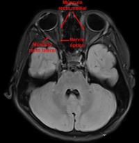
the trochlear and external oculomotor nerves are less frequently affected. Complete knowledge of the cranial nerve course, MRI correlation, and clinical features of cranial nerve palsy is important for radiologists and neurologists to evaluate patients thoroughly. Cranial nerve imaging is usually performed using thin section T2-weighted images based on gradient echo imaging or turbo spin echo sequences, which show the cranial nerves as dark linear structures in contrast to the high signal intensity of the surrounding cerebrospinal fluid. In this article, we review the radiological findings of congenital fibrosis of the extraocular muscles based on brain anatomy and highresolution magnetic resonance imaging.
Downloads
References
Razek H, Maher MA, Kasem M, Helmy E. Imaging of congenital cranial dysinnervation disorders: What radiologist wants to know? Clin Imag. 2021;71:106-16. https://doi.org/10.1016/j.clinimag.2020.10.055
Price JM, Boparai RS, Wasserman BN. Congenital fibrosis of the extraocular muscles: review of recent literatura. Curr Opin Ophthalmol. 2019;30:314-8. https://doi.org/10.1097/ICU.0000000000000592
Miranda MA, Kuschel RC, Miranda GM, Fuentes GA. Anatomía radiológica de la base de cráneo y los nervios craneales parte 2: Nervios craneales. Rev. chil. radiol. [Internet]. 2020 [citado: 2023 mar 19];26(2):62-71. Disponible en: http://www.scielo.cl/scielo.php?script=sci_arttext&pid=S0717-93082020000200062&lng=es
http://dx.doi.org/10.4067/S0717-93082020000200062.
Agarwal V, Vyas S, Dhawan S, Sankhyan N. Isolated congenital absence of cranial nerves: report of two cases. Neuropediatrics. 2018;49(06):405-7. https://doi.org/10.1055/s-0038-1669923.
Kim JH, Hwang JM. Imaging of cranial nerves III, IV, VI in congenital cranial dysinnervation disorders. Korean J Ophthalmol. 2017;31(3):183-93. https://doi.org/10.3341/kjo.2017.0024.
Vivian AJ. Congenital fibrosis of the extra-ocular muscles (CFEOM) and the cranial dysinnervation disorders. Eye (Lond). 2020;34(2):251-5. https://doi.org/10.1038/s41433-019-0700-z.
Downloads
Published
How to Cite
Issue
Section
License
Copyright (c) 2023 Revista Colombiana de Radiología

This work is licensed under a Creative Commons Attribution-NonCommercial-ShareAlike 4.0 International License.
La Revista Colombiana de Radiología es de acceso abierto y todos sus artículos se encuentran libre y completamente disponibles en línea para todo público sin costo alguno.
Los derechos patrimoniales de autor de los textos y de las imágenes del artículo como han sido transferidos pertenecen a la Asociación Colombiana de Radiología (ACR). Por tanto para su reproducción es necesario solicitar permisos y se debe hacer referencia al artículo de la Revista Colombiana de Radiología en las presentaciones o artículos nuevos donde se incluyan.


