
Hyperhomocysteinemia secondary to cystathionine beta-synthetase deficiency. Presentation of an atypical case
DOI:
https://doi.org/10.53903/01212095.198Keywords:
Homocystinuria, Leukoencephalopathies, Magnetic resonance imagingAbstract
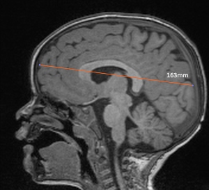
We describe the case of a 2-year-old boy with neurodevelopmental delay, in whom brain MRI showed diffuse white matter changes, diffusion restriction, and hyperintensity of the medial tegmental tract, prior to the diagnosis of hyperhomocysteinemia secondary to Cystathionine beta-synthase deficiency (classic homocystinuria). These findings are not typically described in this disorder, so its diagnosis may be delayed. This case illustrates the need to consider classical homocystinuria in the differential diagnosis of childhood white matter diseases, as well as within the causes of central tegmental tract hyperintensity.
Downloads
References
Garel C, Cont I, Alberti C, Josserand E, Moutard ML, Ducou le Pointe H. Biometry of the corpus callosum in children: MR imaging reference data. AJNR Am J Neuroradiol.2011;32(8):1436-43. doi: https://doi.org/10.3174/ajnr.A2542
Yoshida S, Hayakawa K, Yamamoto A, Aida N, Okano S, Matsushita H, et al. Symmetrical central tegmentario tract (CTT) hyperintense lesions on magnetic resonance imaging in children. Eur Radiol. 200919(2):462-9. doi: https://doi.org/10.1007/s00330-008-1167-7
Aguilera-Albesa S, Poretti A, Honnef D, Aktas M, Yoldi-Petri ME, Huisman TA, et al. T2 hyperintense signal of the central tegmentario tracts in children: disease or normal maturational process? Neuroradiology. 2012;54(8):863-71. doi: https://doi.org/10.1007/s00234-012-1006-z
Barkovich AJ Raybaud C. Pediatric neuroimaging. Sixth ed. Philadelphia: Wolters Kluwer; 2019.
Bublil EM, Majtan T. Classical homocystinuria: From cystathionine beta-synthase deficiency to novel enzyme therapies. Biochimie. 2020;173:48-56. doi: https://doi.org/10.1016/j.biochi.2019.12.007
Weber Hoss GR, Sperb-Ludwig F, Schwartz IVD, Blom HJ. Classical homocystinuria: A common inborn error of metabolism? An epidemiological study based on genetic databases. Mol Genet Genomic Med. 2020;8(6):e1214. doi: https://doi.org/10.1002/mgg3.1214
Sacharow SJ, Picker JD, Levy HL. homocystinuria caused by cystathionine betasynthase deficiency. En: Adam MP, Everman DB, Mirzaa GM, et al., editors. GeneReviewsÆ [Internet]. Seattle (WA): University of Washington, Seattle; 1993-2022.
Morris AA, Kožich V, Santra S, Andria G, Ben-Omran TI, Chakrapani AB, et al. Guidelines for the diagnosis and management of cystathionine beta-synthase deficiency. JInherit Metab Dis. 2017;40(1):49-74. doi: https://doi.org/10.1007/s10545-016-9979-0
Rahman M, Sharma M, Aggarwal P, Singla S, Jain N. Homocystinuria and ocular complications - A review. Indian J Ophthalmol. 2022;70(7):2272-8. doi: https://doi.org/10.4103/ijo.IJO_309_22
Lindstrom K, Ficicioglu C, Kaplan P, Freehauf CL, Levine MA. Low bone mineral density is a common finding in patients with homocystinuria. Mol Genet Metab.2016;117:351-4.
Diaz-Arrastia R. Homocysteine and Neurologic Disease. Arch Neurol.2000;57(10):1422-7. doi: https://doi.org/10.1001/archneur.57.10.1422
Stephan JL. Inaugural cerebral sinovenous thrombosis revealing homocystinuria in a 2-year-old boy. J Child Neurol. 2015;30:107-12.
Karaca M, Hismi B, Ozqul RK, Karaca S, Yilmaz DY, Coskun T, et al. High prevalence of cerebral venous sinus thombosis (CVST) as presentation of cystathionine beta-synthase deficiency in childhood: molecular and clinical findings of Turkish probands. Gene. 2014;534:197-203.
Reddy N, Calloni SF, Vernon HJ, Boltshauser E, Huisman TAGM, Soares BP. Neuroimaging findings of organic acidemias and aminoacidopathies. Radiographics. 2018;38(3):912-31. doi: https://doi.org/10.1148/rg.2018170042
Li CQ, Barshop BA, Feigenbaum A, Khanna PC. Brain magnetic resonance imaging findings in poorly controlled homocystinuria. J Radiol Case Rep. 2018;12(1):1-8. doi: https://doi.org/10.3941/jrcr.v12i1.3207
Knaap MS van der., Valk J, Barkhof F. Magnetic resonance of myelination and myelin disorders. 3rd ed. Springer; 2005.
Brenton JN, Matsumoto JA, Rust RS, Wilson WG. White matter changes in an untreated, newly diagnosed case of classical homocystinuria. J Child Neurol.2014;29(1):88-92. doi: https://doi.org/10.1177/0883073812465012
Ismayilova N, MacKinnon AD, Mundy H, Fallon P. Reversible cerebral white matter abnormalities in homocystinuria. JIMD Rep. 2019;44:115-9. doi: https://doi.org/10.1007/8904_2018_135
Watkins D, Rosenblatt DS. Inherited disorders of folate and cobalamin transport and metabolism. En: Valle D, Beaudet AL, Vogelstein B, Kinzler KW, Antonarakis SE, Ballabio A, Gibson K, Mitchell G, eds. The online metabolic and molecular bases of inherited disease (OMMBID). New York, NY: McGraw-Hill; 2014. Cap. 155.
Gerrard A, Dawson C. Homocystinuria diagnosis and management: it is not all classical. J Clin Pathol. 2022. doi: https://doi.org/10.1136/jcp-2021-208029
Majtan T, Kožich V, Kruger WD. Recent therapeutic approaches to cystathionine beta-synthase-deficient homocystinuria. Br J Pharmacol. 2022. doi: https://doi.org/10.1111/bph.15991
Downloads
Published
How to Cite
Issue
Section
License
Copyright (c) 2023 Revista Colombiana de Radiología

This work is licensed under a Creative Commons Attribution-NonCommercial-ShareAlike 4.0 International License.
La Revista Colombiana de Radiología es de acceso abierto y todos sus artículos se encuentran libre y completamente disponibles en línea para todo público sin costo alguno.
Los derechos patrimoniales de autor de los textos y de las imágenes del artículo como han sido transferidos pertenecen a la Asociación Colombiana de Radiología (ACR). Por tanto para su reproducción es necesario solicitar permisos y se debe hacer referencia al artículo de la Revista Colombiana de Radiología en las presentaciones o artículos nuevos donde se incluyan.


