
Primary leptomeningea presentation of tumors with diffusion restriction. Report of two cases
DOI:
https://doi.org/10.53903/01212095.199Keywords:
Neoplasias del sistema nervioso central, Imagen por resonancia magnética, PediatríaAbstract
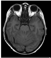
Primary leptomeningeal tumors in pediatrics are uncommon entities. They mostly consist of diffuse glioneuronal tumors, although a few cases of embryonal tumors have also been reported. The complexity in diagnosing this presentation is due to atypical clinical manifestations and difficulties in the differential diagnosis. Diffusion-weighted imaging (DWI) is an imaging modality that is extremely sensitive in detecting water movement in the extracellular space . In neuro-oncology, its utility lies in differentiating between tumors with low cellular density and those with high cellular density, particularly those composed of “small, round, and blue” cells. In this study, we present two cases of disseminated primary leptomeningeal tumors of embryonal origin without a primary brain mass, showing restriction on DWI (hypercellularity). The cases, studied at Garrahan Hospital in the last 3 years, along with a literature review, indicate that the most common imaging finding is diffuse intracranial and intra-spinal leptomeningeal nodular thickening and enhancement. However, no reports were found on the utility of DWI in diagnosing these entities. The article analyzes neuroimaging approaches and diagnostic confirmation to provide opportunities for effective treatment of these diseases in clinical practice.
Downloads
References
Louis DN, Perry A, Wesseling P, et al. The 2021 WHO Classification of Tumors of the Central Nervous System: a summary. Neuro Oncol; 2021;23(8):1231-51. https://doi.org/10.1093/neuonc/noab1063
Osborn AG, Louis DN, Poussaint TY, et al. The 2021 World Health Organization Classification of Tumors of the Central Nervous System: What neuroradiologists need to know. AJNR Am J Neuroradiol. 2022;43(7):928-37. https://doi.org/10.3174/ajnr.A7462
Min L, Yuhao D, Wangming Z. Molecular determinants of medulloblastoma metastasis and leptomeningeal dissemination. Mol Cancer Res. 2021;19:743-52. https://doi.org/10.1158/1541-7786.MCR-20-1026
Ríos CI, De Jesus O. Primitive neuroectodermal tumor. StatPearls [Internet]. Treasure Island (FL): StatPearls Publishing; 2022. PMID: 32965836.
Tanaka H, Yamamoto D, Ikeda M, et al. Embryonal brain tumor with unknown primary lesion and massive cerebrospinal fluid dissemination: A case report. J Clin Neurosci. 2018;54:125-8. https://doi.org/10.1016/j.jocn.2018.04.046
Shih R, Koeller K. Embryonal tumors of the central nervous system: From the radiologic pathology archives. RadioGraphics. 2018;38:2:525-41. https://doi.org/10.1148/rg.2018170182
Morgacheva D, Daks A, Smirnova A, et al. Primary leptomeningeal medulloblastoma in a child: Clinical case report and literature review. Front Pediatr. 2022;10:925340. https://doi.org/10.3389/fped.2022.925340
Ferrara M, Bizzozero L, Fiumara E, et al. “Primary” leptomeningeal dissemination of medulloblastoma. Report of an unusual case. J Neurosurg Sci. 1989;33:219-23. PMID: 2795197.
Suman R, Santosh V, Anandh BA. Primary leptomeningeal medulloblastoma. Pediatr Neurosurg. 2007;43:544-5. https://doi.org/10.1159/000108806
Mehta RI, Cutler AR, Lasky III JL, et al. “Primary” leptomeningeal medulloblastoma. Hum Pathol. 2009;40:1661-5. https://doi.org/10.1016/j.humpath.2009.04.024
Ghosh A, Slopis J, Koenig MK. Primary leptomeningeal medulloblastoma: a rare presentation. AAN Ent. 2018;6:105.
Sublett J, Davenport C, Eisenbrock H, et al. Pediatric primary difusse leptomeningeal Primitive Neuroectodermal Tumor: A case report and literature review. Pediatr Neurosurg. 2017;52(2):114-21. https://doi.org/10.1159/000452807
Tomomasa R, Nakata S, Nobusawa S, et al. Primary diffuse leptomeningeal atypical teratoid/rhabdoid tumor diagnosed by cerebrospinal fluid cytology: case report with molecular genetic analysis. Hum Pathol. 2018;77:116-20. https://doi.org/10.1016/j.humpath.2017.12.026
El-Nabbout B, Shbarou R, Glasier CM, et al. Primary diffuse cerebral leptomeningeal atypical teratoid rhabdoid tumor: report of the first case. J Neurooncol. 2010;98:431-4. https://doi.org/10.1007/s11060-009-0094-z
Gauvain KM, Durham BH, MuHugh M, et al. Rapidly progressive primary leptomeningeal Atypical teratoid/rhabdoid tumor: a report of 2 cases. J Child Neurol. 2012;27:1596-601. https://doi.org/10.1177/0883073812436878
Livermore LJ, Dabbous B, Hofer M, et al. Primary diffuse leptomeningeal atypical teratoid/rhabdoid tumour in an adolescent. Clin Neurol Neurosurg. 2013;115:2170-3. https://doi.org/10.1016/j.clineuro.2013.05.036
Surov A, Meyer HJ, Wienke A. Correlation between apparent diffusion coefficient (ADC) and cellularity is different in several tumors: a meta-analysis. Oncotarget. 2017;8(35):59492-9. https://doi.org/10.18632/oncotarget.17752
Rumboldt Z, Camacho DL, Lake D, et al. Apparent diffusion coefficients for differentiation of cerebellar tumors in children. AJNR Am J Neuroradiol. 2006;27(6):1362-9. PMID: 16775298; PMCID: PMC8133915.
Smirniotopoulos JG, Murphy FM, Rushing EJ, et al. Patterns of contrast enhancement in the brain and meninges. RadioGraphics. 2007;27(2):525-51. https://doi.org/10.1148/rg.272065155
Gardiman MP, Fassan M, Orvieto E, et al. Diffuse leptomeningeal glioneuronal tumors: a new entity? Brain Pathol. 2010;20(2):361-6. https://doi.org/10.1111/j.1750-3639.2009.00285
Lakhani DA, Mankad K, Chhabda S, et al. Diffuse leptomeningeal glioneuronal tumor of childhood. AJNR Am J Neuroradiol. 2020;41(11):2155-9. https://doi.org/10.3174/ajnr.A6737
Peer S, Murumkar V, Kulanthaivelu K, et al. Diffuse leptomeningeal glioneuronal tumor with high-grade features masquerading as tubercular meningitis—a case report. Egypt J Radiol Nucl Med. 2021;52:146. https://doi.org/10.1186/s43055-021-00522-0
Amer EM, Youssef AF, Romeih MA, et al. Role of magnetic resonance imaging in characterization of central nervous system lesions in pediatric patients with leukemia and post-treatment complications. Egypt
Downloads
Published
How to Cite
Issue
Section
License
Copyright (c) 2024 Revista Colombiana de Radiología

This work is licensed under a Creative Commons Attribution-NonCommercial-ShareAlike 4.0 International License.
La Revista Colombiana de Radiología es de acceso abierto y todos sus artículos se encuentran libre y completamente disponibles en línea para todo público sin costo alguno.
Los derechos patrimoniales de autor de los textos y de las imágenes del artículo como han sido transferidos pertenecen a la Asociación Colombiana de Radiología (ACR). Por tanto para su reproducción es necesario solicitar permisos y se debe hacer referencia al artículo de la Revista Colombiana de Radiología en las presentaciones o artículos nuevos donde se incluyan.


