
Moyamoya syndrome, from image to diagnosis. Presentation of a case
DOI:
https://doi.org/10.53903/01212095.232Keywords:
Enfermedad de moyamoya, Trastornos cerebrovasculares, Enfermedades arteriales cerebralesAbstract
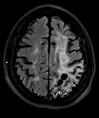
Moyamoya disease and Moyamoya syndrome refer to a progressive steno-occlusive vasculopathy in the terminal portions of the bilateral internal carotid arteries and/or the proximal parts of the middle and anterior cerebral arteries, with prominent formation of collateral arteries. Being able to generate irreversible damage to cerebral hemodynamics due to the progressive nature of the affected cerebral vessels, the pathological examination does not show atherosclerotic or inflammatory lesions and the cause of the stenosis is the overgrowth of the smooth muscle layer, with thrombotic changes.
The disease leads not only to a different degree of stenosis and occlusions of the great arteries in the anterior part of the circle of Willis, but also to the development of collateral vasculature that produces a typical angiographic image, termed 'puffs of smoke' or 'puff'. of cigarette smoke'.
We present the case of a 28-year-old female patient who presents with insidious symptoms and a previous wrong clinical and imaging diagnosis, in whom a follow-up magnetic resonance of the brain was performed, evidencing findings by imaging and angioresonance compatible with Moyamoya disease; the imaging findings of the syndrome in the different modalities of radiological studies are analyzed.
Downloads
References
Li J, Jin M, Sun X, et al. Imaging of moyamoya disease and moyamoya syndrome: Current status. J Comput Assist Tomogr. 2019;43(2):257-63. https://doi.org/10.1097/RCT.0000000000000834.
Scott RM, Smith ER. Moyamoya disease and moyamoya syndrome. N Engl J Med.2009;360(12):1226-37.
Lim M, Cheshier S, Steinberg G. New vessel formation in the central nervous system during tumor growth, vascular malformations, and Moyamoya. Curr Neurovasc Res.2006;3(3):237-45.
Hasuo K, Mihara F, Matsushima T. MRI and MR angiography in moyamoya disease. J Magn Reson Imaging. 1998;8(4):762-6.
Piao R, Oku N, Kitagawa K, et al. Cerebral hemodynamics and metabolism in adult moyamoya disease: comparison of angiographic collateral circulation. Ann Nucl Med. 2004;18(2):115-21.
Tarasów E, Kułakowska A, Lukasiewicz A, et al. Moyamoya disease: Diagnostic imaging. Pol J Radiol. 2011;76(1):73-9.
Kuroda S, Houkin K. Moyamoya disease: Current concepts and future perspectives. Lancet Neurology. 2008;7(11):1056-66. https://doi.org/10.1016/S14744422(08)70240-0
Miyamoto S, Yoshimoto T, Hashimoto N, et al. Effects of extracranial-intracranial bypass for patients with hemorrhagic Moyamoya disease: Results of the Japan Adult Moyamoya Trial. Stroke. 2014;45(5):1415-21. https://doi.org/10.1161/STROKEAHA.113.004386
Downloads
Published
How to Cite
Issue
Section
License
Copyright (c) 2024 Revista Colombiana de Radiología

This work is licensed under a Creative Commons Attribution-NonCommercial-ShareAlike 4.0 International License.
La Revista Colombiana de Radiología es de acceso abierto y todos sus artículos se encuentran libre y completamente disponibles en línea para todo público sin costo alguno.
Los derechos patrimoniales de autor de los textos y de las imágenes del artículo como han sido transferidos pertenecen a la Asociación Colombiana de Radiología (ACR). Por tanto para su reproducción es necesario solicitar permisos y se debe hacer referencia al artículo de la Revista Colombiana de Radiología en las presentaciones o artículos nuevos donde se incluyan.


