
Concordance in the measurement of the distance between the anterior tibial tuberosity to the trochlear groove in axial tomography of the patella at 20 degrees of flexion with respect to 0 degrees
DOI:
https://doi.org/10.53903/01212095.238Keywords:
Patellofemoral pain syndrome, Abdominal pain, Computed axial tomographyAbstract
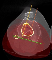
Background: Patellofemoral malalignment is a frequent pathology in young people. With an early diagnosis, a timely intervention can be made, so the distance between the intertrochlear Groove and the anterior tibial tuberosity (ICG-ATT) is essential to indicate which patients need surgery. Currently patellar axial tomography is taken with cuts at 0, 20 and 45 degrees of flexion, although there is not enough information in the literature that specifies and justifies which degrees of flexion should be used, the purpose of the study is to establish the protocol for patellar axial tomography. Objective: To establish whether there is agreement between the ICG-ATT measurement performed at 20 and 0 degrees of flexion. Methods: A concordance study was carried out, 90 patients were taken for a total of 180 knees, and ICG-ATT measurement performed on these images, interposing cuts at 0 degrees and 20 degrees of flexion, to evaluate if there is agreement between the two measurements. Results: There is an almost perfect agreement of 0.909 between the measurement at 0 and 20 degrees of flexion on the left side and an almost perfect agreement of 0.8872 between the measurement at 0 and 20 degrees of flexion on the right side of the images. evaluated. Conclusions: Near perfect agreement was found between the ICG-ATT measurement performed at 0 and 20 degrees of flexion, meaning that there is no difference in taking the measurement at these degrees. We propose that patellar CT protocols should be taken at 20 degrees, supported by the biomechanics of the knee, because the stability of flexion at 20 degrees is based primarily on bone restriction as well as decreased radiation dose. We suggest future research with standardized protocols at 20 degrees of flexion.
Downloads
References
Boling M, Padua D, Marshall S, et al. Gender differences in the incidence and prevalence of patellofemoral pain syndrome. Scand J Medsci Sports 2010: 725-730. Available from: https://doi.org/10.1111/j.1600-0838.2009.00996.x
Drew BT, Redmond AC, Smith TO, et al. Which patellofemoral joint imaging features are associated with patellofemoral pain? Systematic review and meta-analysis. Osteoarthr Cartil [Internet]. 2016;24(2):224–36. Available from: http://dx.doi.org/10.1016/j.joca.2015.09.004
Gulati A, Mcelrath C, Madhwa V, et al. current clinical, radiological and treatment perspectives of patellofemoral pain syndrome. Br J radiol;91:2017.
Loudon J, biomechanics and pathomechanics of the patellofemoral joint. Int Sports Phys Ther 2016 Dec 11(6) 820-830
Sherman SL, Raines BT, Burch MB, et al. Patellofemoral imaging and analysis. Oper Tech Sports Med [Internet]. 2019;150684. Available from: https://doi.org/10.1016/j.otsm.2019.150684
Laugharne E, Bali N, Puroshothamdas S, et al. Variability of Measurement of patellofemoral indices with knee flexion and quadricpes contraction: An MRI-based anatomical study. Knee Surg Relat Res 2016; 28, 297-301.
Worden A, Kaar S, Owen J, Cutuk A, et al. Radiographic and Anatomic Evaluation of Tibial Tubercle to Trochlear Groove Distance. J Knee Surg. 2016;29(7):589–93.
Mayo W, Hara A. Mahesh M. et al, How I do it: Managing radiation dose in CT. Radiology: volume 273: Number 3- December 2014.
Biswas D, Bible J. Bohan M, et al. Radiation exposure from musculoskeletal computerized tomographic scans. J bone Joint Surg Am. 2009.
Barzan M. Maine S. Modenese L, et al. Patellofemoral joint alignment is a major risk factor for recurrent patellar dislocation in children and adolescents: a systematic review. JISAKOS 20, 3:287-297. Doi: 10.1136/jisakos-2017-000189.
Sharon S, Ying B, Jason K, et al. The difference between computed tomography and magnetic resonance imaging measurements of tibial tubercle-trochlear groove distance for patients with or without patellofemoral instability: A systematic review and metaanalysis. J knee surg. 2020 Aug;33(8):768–776.
Schoettle P, Zanetti M, Seifert B, et al. The tibial tuberosity-trochlear groove distance; a comparative study between CT an MRI scanning. The knee. 2006;13(1):26-31.
Suomalainen J, Regalado G, Joukainen A. et al. Effects of knee flexion and extension on the tibial tuberosity-trochlear groove (TT-TG) distance in adolescents. Journal of experimentatal orthopaedics. 2018. https://doi.org/10.1186/s40634-018-0149-1
Miyanishi K, Nagamine R, Murayama S, et al. Tibial tubercle malposition in patellar joint instability: A computed tomography study in full extension and at 30 flexion. Acta orthopaedoca. 2000. DOI: 10.1080/000164700317411898.
Fithian D, Neyret P, Elvire S. Patellar instability: The Lyon experience. Current orthopaedic practice. 2008;19(3):328-338.
Lin Ll, A concordance correlacion coefficient to evaluate reproducibility. Biometrics 1989 Mar, 255-268.
Figueroa D, Novoa F, Melean P, et al. Utilidad de la resonancia magnética en la evaluación del mal alineamiento patelar. Rev . Esp Cir Ortop Traumatol. 2014;58(1):19-23
Tecklenburg K, Feller J, Whiteheead T, et al. Outcome of surgery for recurrent patellar dislocation base don the distance of the tibial tuberosity to the trochlear groove. J Bone Joint Surg 2010;92-B:1376-80.
Marquez L, Andersen J, Lenchik L, et al. Variability in patelofemoral alignment measurements on MRI influence of knee position. AJR 208, May 2017
Dietrich T, Betz M, Pfirmann C, et al. End stage extension of the knee and its influence on tibial tuberosity trochlear groove distance (TTTG) in asymptomatic volunteers. Knee surg sports traumatol arthrosc 2014 22:214-218
Downloads
Published
How to Cite
Issue
Section
License
Copyright (c) 2024 Revista Colombiana de Radiología

This work is licensed under a Creative Commons Attribution-NonCommercial-ShareAlike 4.0 International License.
La Revista Colombiana de Radiología es de acceso abierto y todos sus artículos se encuentran libre y completamente disponibles en línea para todo público sin costo alguno.
Los derechos patrimoniales de autor de los textos y de las imágenes del artículo como han sido transferidos pertenecen a la Asociación Colombiana de Radiología (ACR). Por tanto para su reproducción es necesario solicitar permisos y se debe hacer referencia al artículo de la Revista Colombiana de Radiología en las presentaciones o artículos nuevos donde se incluyan.


