
Contrast-enhanced mammography: imaging patterns of breast cancer molecular subtypes
DOI:
https://doi.org/10.53903/01212095.257Keywords:
Breast cancer, Mammography, inmuno-histoquimica, Intrisec subtipesAbstract
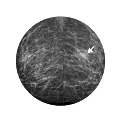
Introduction: Breast cancer (BC) is a heterogeneous disease, classified into three main intrinsic subtypes: hormone receptor positive (HR+), HER-2 oncogene overexpressing and triple negative (TN), each with representative imaging findings. Objective: Characterize and classify the findings of intrinsic subtypes of breast cancer using contrast-enhanced mammography imaging.
Methodology: A descriptive analysis was conducted on a subsample of patients who were enrolled in a specialized diagnostic care program for breast pathology. From 2019 to 2022, clinical findings, histology, immunohistochemistry results and CEM (contrast-enhanced mammography) features were recorded from 71 patients with BC. Results: 49 cases of breast carcinoma that underwent CEM were hormone receptor positive, 3 had HER-2 enriched and 19 were triple-negative (TN). Spiculated tumors were associated with hormone receptor positive, calcifications occurred in hormone receptor positive and HER2 enriched, being more frequent in HER2 enriched breast cancer, and ring enhancement, was associated with the TN subtype. Conclusion: In the age of minimally invasive medicine, the use of contrast-enhanced mammography images to forecast intrinsic subtypes of breast cancer is an innovative and auspicious development. Nevertheless, gene expression analysis will continue to be the gold standard for the time being.
Downloads
References
Sung H, Ferlay J, Siegel RL, Laversanne M, Soerjomataram I, Jemal A, et al. Global Cancer Statistics 2020: GLOBOCAN estimates of incidence and mortality worldwide for 36 cancers in 185 countries. CA Cancer J Clin. 2021;71(3):209-49
Sørlie T, Perou CM, Tibshirani R, Aas T, Geisler S, Johnsen H, et al. Gene expression patterns of breast carcinomas distinguish tumor subclasses with clinical implications. Proc Natl Acad Sci U S A. 2001;98(19):10869-74
Romond EH, Pérez EA, Bryant J, Suman VJ, Geyer CE, Davidson NE, et al. Trastuzumab plus adjuvant chemotherapy for operable HER2-positive breast cancer. N Engl J Med. 2005;353(16):1673-84
Foulkes WD, Smith IE, Reis-Filho JS. Triple-negative breast cancer. N Engl J Med. 2010;363(20):1938-48.
Perou CM, Sørlie T, Eisen MB, van de Rijn M, Jeffrey SS, Rees CA, et al. Molecular portraits of human breast tumors. Nature. 2000;406(6797):747-52.
Dent R, Trudeau M, Pritchard KI, Hanna WM, Kahn HK, Sawka CA, et al. Triplenegative breast cancer: clinical features and patterns of recurrence. Clin Cancer Res. 2007;13(15 Pt 1):4429-34.
Collett K, Stefansson IM, Eide J, Braaten A, Wang H, Eide GE, et al. A basal epithelial phenotype is more frequent in interval breast cancers compared with screen detected tumors. Cancer Epidemiol Biomarkers Prev. 2005;14(5):1108-12.
Pal SK, Childs BH, Pegram M. Triple negative breast cancer: unmet medical needs. Breast Cancer Res Treat. 2011;125(3):627-36.
Lee CH, Philips J, Sung JS, Lewin JM, Newell MS. Contrast Enhanced Mammography (CEM). A supplement to ACR BI-RADS Mammography 2013. American College of Radiology. 2022.
Li N, Gong W, Xie Y, Sheng L. Correlation between the CEM imaging characteristics and different molecular subtypes of breast cancer. Breast. 2023;72:103595.
Seo BK, Pisano ED, Kuzimak CM, Koomen M, Pavic D, Lee Y, et al. Correlation of HER-2/neu overexpression with mammography and age distribution in primary breast carcinomas. Acad Radiol. 2006;13(10):1211-8.
Wang S, Wang Z, Li R, You C, Mao N, Jiang T, et al. Association between quantitative and qualitative image features of contrast-enhanced mammography and molecular subtypes of breast cancer. Quant Imaging Med Surg. 2022;12(2):1270-80.
Fallenberg EM, Dromain C, Diekmann F, Engelken F, Krohn M, Singh JM, et al. Contrast-enhanced spectral mammography versus MRI: Initial results in the detection of breast cancer and assessment of tumour size. Eur Radiol. 2014;24(1):256-64.
Łuczyńska E, Heinze-Paluchowska S, Hendrick E, Dyczek S, Ryś J, Herman K, et al. Comparison between breast MRI and contrast-enhanced spectral mammography. Med Sci Monit. 2015;21:1358-67.
Xiang W, Rao H, Zhou L. A meta-analysis of contrast-enhanced spectral mammography versus MRI in the diagnosis of breast cancer. Thorac Cancer. 2020;11(6):1423-32.
Uematsu T, Kasami M, Yuen S. Triple-negative breast cancer: correlation between MR imaging and pathologic findings. Radiology. 2009;250(3):638-47.
Downloads
Published
How to Cite
Issue
Section
License
Copyright (c) 2024 Revista Colombiana de Radiología

This work is licensed under a Creative Commons Attribution-NonCommercial-ShareAlike 4.0 International License.
La Revista Colombiana de Radiología es de acceso abierto y todos sus artículos se encuentran libre y completamente disponibles en línea para todo público sin costo alguno.
Los derechos patrimoniales de autor de los textos y de las imágenes del artículo como han sido transferidos pertenecen a la Asociación Colombiana de Radiología (ACR). Por tanto para su reproducción es necesario solicitar permisos y se debe hacer referencia al artículo de la Revista Colombiana de Radiología en las presentaciones o artículos nuevos donde se incluyan.


