
Myxoid liposarcoma with unusual clinical presentation: imaging diagnosis and histopathologic correlation. A case report
DOI:
https://doi.org/10.53903/01212095.268Keywords:
Liposarcoma, Liposarcoma, myxoid, Magnetic resonance imagingAbstract
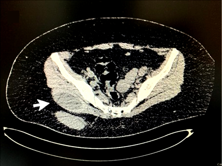
Myxoid liposarcoma is the second most frequent malignant neoplasm of all types of liposarcoma, usually located in the lower limbs and retroperitoneum. It is characterized as a slow-growing, painless soft tissue mass that develops especially in the intermuscular and rarely intramuscular compartments and, depending on its cellularity, can become highly metastatic. Imaging diagnosis and its histopathological correlation is essential to provide timely treatment; however, this can become a great challenge due to the lack of specificity of its findings in first line imaging studies that are generally available in hospitals up to second level. This makes the need for more advanced studies essential, such as magnetic resonance imaging (MRI), where the role of the radiologist is
essential for the detection of subtle but pathognomonic findings of this pathology. We present the case of a young adult patient with an unusual presentation of a myxoid liposarcoma who presents with pain and mass at the level of the right buttock; her imaging studies are performed
and analyzed and correlated with histopathological findings.
Downloads
References
Dei Tos AP. Liposarcomas: diagnostic pitfalls and new insights. Histopathology. 2014;64(1):38-52. http://dx.doi.org/10.1111/his.12311
Sbaraglia M, Bellan E, Dei Tos AP. The 2020 WHO Classification of Soft Tissue Tumours: news and perspectives. Pathologica. 2020;113(2):70-84. http://dx.doi.org/10.32074/1591-951x-213
Loubignac F, Bourtoul C, Chapel F. Myxoid liposarcoma: a rare soft-tissue tumor with a misleading benign appearance. World J Surg Oncol. 2009;7(1). http://dx.doi.org/10.1186/1477-7819-7-42
Petscavage-Thomas JM, Walker EA, Logie CI, Clarke LE, Duryea DM, Murphey MD. Soft-tissue myxomatous lesions: Review of salient imaging features with pathologic comparison. Radiographics. 2014;34(4):964-80. http://dx.doi.org/10.1148/rg.344130110
Benjaminov O, Gutman H, Nyabanda R, Keinan R, Sabach G, Levavi H. Myxoid liposarcoma: An unusual presentation. AJR Am J Roentgenol. 2007;188(3):817-21. http://dx.doi.org/10.2214/ajr.05.0246
Benjaminov O, Gutman H, Nyabanda R, Keinan R, Sabach G, Levavi H. Myxoid liposarcoma: An unusual presentation. Am J Roentgenol. 2007;188(3):817-21. https://doi.org/10.2214/AJR.05.0246
Morán LM, Vega J, Gómez-León N, Royuela A. Myxomas and myxoid liposarcomas of the extremities: Our preliminary findings in conventional, perfusion, and diffusion magnetic resonance. Acta Radiol Open. 2022;11(10):20584601221131481. https://doi.org/10.1177/20584601221131481
Chung WJ, Chung HW, Shin MJ, Lee SH, Lee MH, Lee JS, et al. MRI to differentiate benign from malignant soft-tissue tumours of the extremities: a simplified systematic imaging approach using depth, size and heterogeneity of signal intensity. Br J Radiol. 2012;85(1018):e831-6. http://dx.doi.org/10.1259/bjr/27487871
Baheti AD, Tirumani SH, Rosenthal MH, Howard SA, Shinagare AB, Ramaiya NH, et al. Myxoid soft-tissue neoplasms: Comprehensive update of the taxonomy and MRI features. AJR Am J Roentgenol. 2015;204(2):374-85. http://dx.doi.org/10.2214/ajr.14.12888
Tfayli Y, Baydoun A, Naja A, Saghieh S. Management of myxoid liposarcoma of the extremity (Review). Oncol Lett. 2021;22(2). http://dx.doi.org/10.3892/ol.2021.12857
Tuzzato G, Laranga R, Ostetto F, Bubbico E, Vara G, Bianchi G. Primary high-grade myxoid liposarcoma of the extremities: Prognostic factors and metastatic pattern. Cancers. 2022;14(11):2657. https://doi.org/10.3390/cancers14112657
Downloads
Published
How to Cite
Issue
Section
License
Copyright (c) 2025 Revista Colombiana de Radiología

This work is licensed under a Creative Commons Attribution-NonCommercial-ShareAlike 4.0 International License.
La Revista Colombiana de Radiología es de acceso abierto y todos sus artículos se encuentran libre y completamente disponibles en línea para todo público sin costo alguno.
Los derechos patrimoniales de autor de los textos y de las imágenes del artículo como han sido transferidos pertenecen a la Asociación Colombiana de Radiología (ACR). Por tanto para su reproducción es necesario solicitar permisos y se debe hacer referencia al artículo de la Revista Colombiana de Radiología en las presentaciones o artículos nuevos donde se incluyan.


