
Dosimetric evaluation of 2D mammography and tomosynthesis with synthesized images at the Fundación CTIC
DOI:
https://doi.org/10.53903/01212095.301Keywords:
Radiation dose, Mammography, Package inserts for the patientAbstract
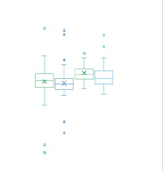
Objective: Given the concern about a higher dose when performing mammography with tomosynthesis and the few publications on comparative radiation doses between 2D mammography and tomosynthesis, we sought to demonstrate that mammography with tomosynthesis and synthesized images does not increase the dose compared to standard 2D digital mammography. Materials and Methods: Retrospective cross-sectional study. Data included patient information, acquisition parameters, entrance dose, and mean glandular dose per projection, differentiating between 2D acquisition and tomosynthesis. Results: A total of 689 mammographic projections were documented, of which 367 were tomosynthesis projections. The doses showed notable similarities between 2D mammography and tomosynthesis techniques. For CC projection, the average dose was 1.48 mGy in 2D acquisition and 1.45 in tomosynthesis (p=0.6) and in MLO, 1.63 mGy and 1.58 mGy respectively (p=0.09). Conclusions: Dosimetric values in tomosynthesis mammography were slightly lower than those in conventional mammography, highlighting the capacity to synthesize 2D images. The implementation of synthesized images is proposed as a strategy to optimize the radiation dose in mammographic studies, improving safety and efficiency in clinical practice.
Downloads
References
Rafferty EA, Park JM, Philpotts LE, Poplack SP, Sumkin JH, Halpern EF, et al. Diagnostic accuracy and recall rates for digital mammography and digital mammography combined with one-view and two-view tomosynthesis: Results of an enriched reader
study. Am J Roentgenol. 2014;202(2):273-81.
Gilbert FJ, Tucker L, Gillan MGC, Willsher P, Cooke J, Duncan KA, et al. Accuracy of digital breast tomosynthesis for depicting breast cancer subgroups in a UK retrospective reading study (TOMMY Trial). Radiology. 2015;277(3):697-706.
Houssami N. Evidence on synthesized two-dimensional mammography versus digital mammography when using tomosynthesis (three-dimensional mammography) for population breast cancer screening. Clin Breast Cancer. 2018;18(4):255-60.e1.
ICRP. Diagnostic reference levels in medical imaging. ICRP Publication 135. Ann. ICRP. 2017;46(1).
Feng SSJ, Sechopoulos I. Clinical digital breast tomosynthesis system: Dosimetric characterization. Radiology. 2012;263(1):35-42.
James JR, Pavlicek W, Hanson JA, Boltz TF, Patel BK. Breast radiation dose with CESM compared with 2D FFDM and 3D tomosynthesis mammography. Am J Roentgenol. 2017;208(2):362-72.
Gennaro G, Bernardi D, Houssami N. Radiation dose with digital breast tomosynthesis compared to digital mammography: per-view analysis. Eur Radiol. 2018;28(2):573-81.
Skaane P, Bandos AI, Gullien R, Eben EB, Ekseth U, Haakenaasen U, et al. Comparison of digital mammography alone and digital mammography plus tomosynthesis in a population-based screening program. Radiology. 2013;267(1):47-56.
Díaz Henao F. Determinación de niveles de referencia en procedimientos de mamografía digital tomosíntesis [Tesis] Universidad Nacional de Colombia [internet]. 2020 May 23 [citado: 2023 may 15]. Disponible en: https://repositorio.unal.edu.co/handle/unal/77555
Suleiman M. Diagnostic reference levels for digital mammography in Australia [Tesis]. The University of Sydney [internet]. 2018 [citado: 2023 may 15]. Disponible en: http://hdl.handle.net/2123/18930.
Olgar T, Kahn T, Gosch D. Average glandular dose in digital mammography and breast tomosynthesis. Rofo. 2012;184(10):911-8.
Downloads
Published
How to Cite
Issue
Section
License
Copyright (c) 2025 Revista Colombiana de Radiología

This work is licensed under a Creative Commons Attribution-NonCommercial-ShareAlike 4.0 International License.
La Revista Colombiana de Radiología es de acceso abierto y todos sus artículos se encuentran libre y completamente disponibles en línea para todo público sin costo alguno.
Los derechos patrimoniales de autor de los textos y de las imágenes del artículo como han sido transferidos pertenecen a la Asociación Colombiana de Radiología (ACR). Por tanto para su reproducción es necesario solicitar permisos y se debe hacer referencia al artículo de la Revista Colombiana de Radiología en las presentaciones o artículos nuevos donde se incluyan.


