
Complications associated with the presence of a double collecting system. Presentation of two cases
DOI:
https://doi.org/10.53903/01212095.302Keywords:
Urinary incontinence, Urinary tract infection, Vesicoureteral reflux, Tomography, Urography, UreterAbstract
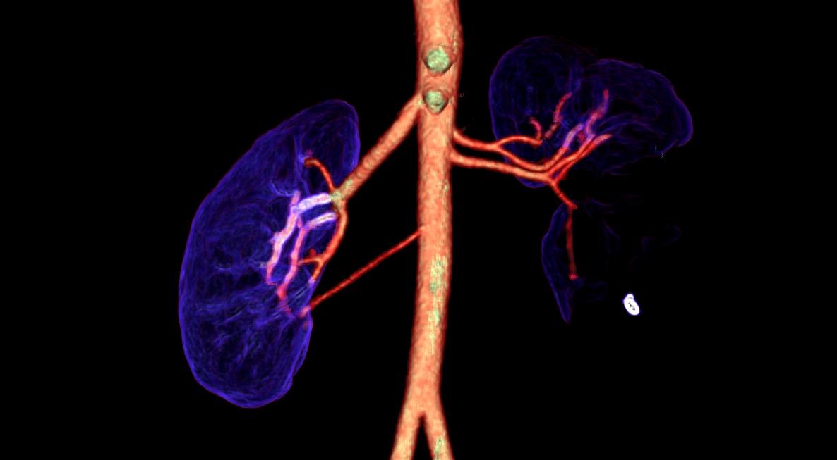
Congenital abnormalities of the upper urinary tract are present in 3-11 % of the population and alone account for up to 50 % of malformations occurring during the embryonic stage. These embryogenesis alterations affecting the genitourinary system are classified according to renal morphology, the anatomical position of the kidney in the abdominopelvic cavity, number (agenesis
and supernumerary kidneys), and abnormalities in the development of the collecting system, the latter being the most prevalent. The double collecting system is the most common alteration, usually asymptomatic, and often an anatomical variant without clinical relevance, typically diagnosed incidentally in most cases. However, in some scenarios, it can lead to significant complications that
may compromise renal function and affect the quality of life of affected patients. Regarding imaging methods, there are many diagnostic aids that can be used to detect these different conditions, and depending on the patient’s age, some diagnostic methods are preferred over others. The
objective of this article is to demonstrate the complications associated with the double collecting system through two clinical cases.
Downloads
References
Houat AP, Guimarães C, Takahashi M, Rodi G, Gasparetto T, Blasbalg R, et al. Congenital anomalies of the upper urinary tract: A comprehensive review. Radiographics. 2021;41(2):462-86. https://doi.org/10.1148/rg.2021200078
Didier R, Chow J, Kwatra N, Retik A, Lebowitz R.. The duplicated collecting system of the urinary tract: embryology, imaging appearances and clinical considerations. Pediatr Radiol. 2017;47(11):1526-38. https://doi.org/10.1007/s00247-017-3904-z
Fernbach SK, Feinstein KA, Spencer K, Lindstrom CA. Ureteral duplication and its complications. Radiographics. 1997;17(1):109-27. https://doi.org/10.1148/radiographics.17.1.9017803
Privett JT, Jeans WD, Roylance J. The incidence and importance of renal duplication. Clin Radiol. 1976;27(4):521-30. https://doi.org/10.1016/s0009-9260(76)80121-3
Amis ES Jr, Cronan JJ, Pfister RC. Lower moiety hydronephrosis in duplicated kidneys. Urology. 1985;26(1):82-8. https://doi.org/10.1016/0090-4295(85)90268-7
Rehder P, Petersen J, Hofmann KJ, Schenk C, Trieb T, Glodny B. Yo-yo reflux in an incomplete duplex system causing severe hydronephrosis in a patient with contralateral renal agenesis. Ren Fail. 2008;30(8):818-21. https://doi.org/10.1080/08860220802272605
Yonli DS, Chakroun M, Zaghbib S, Ye D, Bouzouita A, Derouiche A, Ben Slama MR et al. Bilateral duplex collecting system with bilateral vesicoureteral reflux: a case report. J Med Case Rep. 2019;13(1):128. https://doi.org/10.1186/s13256-019-2058-z
Chionardes MA, Kurniawan Liemarto A, Gunardi SL. Unilateral duplicated collecting system and ureter with severe hydroureteronephrosis and ectopic ureter insertion of upper pole moiety: A case report. Ann Med Surg. 2012;74:103255. https://doi.org/10.1016/j.amsu.2022.103255
Surabhi V, Menias C, George V, Matta E, Kaza R, Hasapes J. MDCT and MR urogram spectrum of congenital anomalies of the kidney and urinary tract diagnosed in adulthood. AJR. Am J Roentgenol. 2015;205(3):W294-304. https://doi.org/10.2214/AJR.14.12867
Melkamu A, Darge K, Dillman JR, Carr M, Epelman M. Magnetic resonance urography in evaluation of duplicated renal collecting systems. Magn Reson Imaging Clin N Am. 2013;21(4):717-30. https://doi.org/10.1016/j.mric.2013.04.002
Potenta SE, D’Agostino R, Sternberg KM, Tatsumi K, Perusse K. CT urography for evaluation of the ureter. Radiographics. 2015;35(3):709-26. https://doi.org/10.1148/rg.2015140209
Cheng K, Cassidy F, Aganovic L, Taddonio M, Vahdat N. CT urography: how to optimize the technique. Abdom Radiol (NY). 2019;44(12):3786-99. https://doi.org/10.1007/s00261-019-02111-2
Kawashima A, Vrtiska TJ, LeRoy AJ, Hartman RP, McCollough CH, King BF Jr. CT urography. Radiographics. 2004;24(Suppl 1):S35-54; discussion S55-8. https://doi.org/10.1148/rg.24si045513
Downloads
Published
How to Cite
Issue
Section
License
Copyright (c) 2025 Revista Colombiana de Radiología

This work is licensed under a Creative Commons Attribution-NonCommercial-ShareAlike 4.0 International License.
La Revista Colombiana de Radiología es de acceso abierto y todos sus artículos se encuentran libre y completamente disponibles en línea para todo público sin costo alguno.
Los derechos patrimoniales de autor de los textos y de las imágenes del artículo como han sido transferidos pertenecen a la Asociación Colombiana de Radiología (ACR). Por tanto para su reproducción es necesario solicitar permisos y se debe hacer referencia al artículo de la Revista Colombiana de Radiología en las presentaciones o artículos nuevos donde se incluyan.


