
Case report of a neuroendocrine neoplasm: findings on ultrasound and angiotomography of a carotid body tumor
DOI:
https://doi.org/10.53903/01212095.305Keywords:
Carotid body, Neuroendocrine neoplasms, Carcinoma transitional cell UltrasonographyAbstract
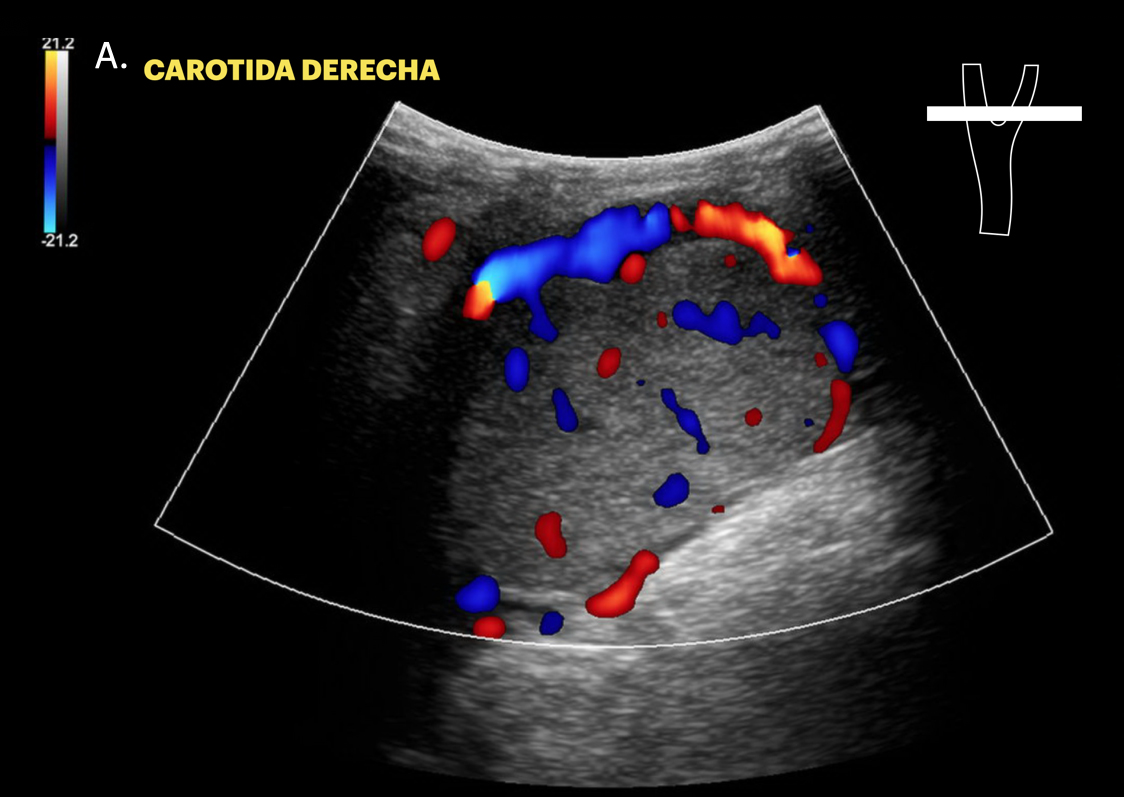
The carotid paraganglioma is an infrequent neuroendocrine tumor originating from the paraganglia of the autonomic system and presents challenges in differential diagnosis due to its low incidence and diversity of clinical presentations. In this article, we will present a case of an 82-year-old woman with varied symptoms and highlight the importance of multidisciplinary management in
the diagnosis and treatment of this pathology. Emphasis is placed on radiological findings, such as ultrasound and computed tomography angiography, which are crucial to confirm the diagnosis. Additonaly, the importance of surgical resection as the treatment of choice is pointed out. In conclusion, a comprehensive and early approach based on clinical evaluation and imaging tools can facilitate the diagnosis and timely management of carotid paraganglioma.
Downloads
References
Lin EP, Chin BB, Fishbein L, Moritani T, Montoya SP, Ellika S, Newlands S. Head and neck paragangliomas: Am updated on the molecular classification, state of the art imaging, and management recommendations. Radiol Imaging Cancer. 2022;4(3):e210088. https://doi.org/10.1148/rycan.210088
Hoang VT, Trinh CT, Lai TAK, Doan DT, Tran TTT. Carotid body tumor: a case report and literature review. J Radiol Case Rep. 2019;13(8):19-30. https://doi.org/10.3941/jrcr.v13i8.3681
Cárdenas K, Zamora G. Radiología en el glomus carotídeo. Presentación de un caso. Imaging in carotid body paraganglioma. A case report. Metro Ciencia. 2021:29(2):63-9. https://doi.org/10.47464/MetroCiencia/vol29/3/2021/63-69
González-Urquijo M, Hinojosa-González D, Viteri-Pérez VH, Llausas-Villarreal A, Becerril-Gaitan A, González-González M, et al. An analysis from the CAPACITY database of outcomes of preoperative embolization before carotid body tumor surgery compared with resection alone. J Vasc Surg. 2023;77(5):1447-52. https://doi.org/10.1016/j.jvs.2023.01.012
Delantoni A, Raftopoulos A, Pol Anagnostaki A, Orhan K. B-mode and color Doppler imaging of carotid paragangliomas in different neck regions. J Ultrason. 2020;20(82):e218-e221. https://doi.org/10.15557/JoU.2020.0037
Alqhtani AJ, Al Qahtani SM, Alharbi MK, Aljehani MH, Alharbi A. Carotid body tumor: a case report of a rare neuroendocrine neoplasm. Cureus. 2023;15(7):e42224. https://doi.org/10.7759/cureus.42224.
Avgerinos N, Avgerinos I, Troupis T, et al. Carotid Body tumor in a 26-year-old male patient managed with preoperative embolization. Curaeus. 2023;15(12):e 49917. https://doi.org/10.7759/cureus.49917
Kihara C, Patel S, Moss R. A rapidly progressing carotid body tumor: a case report. Cureus.2023;15(8): e43654. https://doi.org/10.7759/cureus.43654
Downloads
Published
How to Cite
Issue
Section
License
Copyright (c) 2025 Revista Colombiana de Radiología

This work is licensed under a Creative Commons Attribution-NonCommercial-ShareAlike 4.0 International License.
La Revista Colombiana de Radiología es de acceso abierto y todos sus artículos se encuentran libre y completamente disponibles en línea para todo público sin costo alguno.
Los derechos patrimoniales de autor de los textos y de las imágenes del artículo como han sido transferidos pertenecen a la Asociación Colombiana de Radiología (ACR). Por tanto para su reproducción es necesario solicitar permisos y se debe hacer referencia al artículo de la Revista Colombiana de Radiología en las presentaciones o artículos nuevos donde se incluyan.


