Signs suggestive of osteochondral lesions on knee MRI scans
DOI:
https://doi.org/10.53903/01212095.401Keywords:
Magnetic resonance imaging, Knee, Cartilage, articular, OsteochondritisAbstract
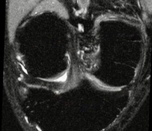
Introduction: The term osteochondral injury or defect is an abnormality of articular cartilage and subchondral bone, which can be caused by a variety of acute and chronic entities. These lesions are manifested in magnetic resonance imaging from discrete changes in signal intensity of the articular cartilage, to loss of morphology associated with bone edema. Objectives: The main objective of this study was to determine the frequency and quantitatively characterize (by edema volume and chondral defect area) osteochondral lesions in symptomatic patients evaluated with knee magnetic resonance imaging. Secondary objectives were to describe the most frequent location of these lesions and to evaluate the statistical association with concomitant pathologies such as meniscal and anterior cruciate ligament lesions. Methodology: Epidemiological and radiological characteristics were retrospectively collected in patients who underwent knee MRI scans at the Radiology Service of a private clinic in Caracas in 2022, identifying suggestive signs of osteochondral lesions in 477 studies. The volume of subchondral bone edema was classified by dividing patients into 3 groups: mild (0-3000 mm3), moderate (between 3001 mm3 and 10000 mm3), and severe (greater than 10000 mm3). Results and Conclusions: Out of a total of 477 knee studies, 229 (48 %) demonstrated the presence of subchondral edema. The age group with the highest frequency of subchondral edema ranged from 41 to 60 years. The patella was found to be the most common site of mild subchondral edema; however, moderate and severe edema was most frequently located in the medial condyle (20.7 %) and the medial tibial plateau (19 %). Cartilage involvement was present in 82.5 % of positive studies. Early diagnosis of osteochondral lesions can help the treating physician slow their progression or even reverse bone edema before the lesion progresses to a severe cartilage defect.
Downloads
References
Chien A, Weaver J, Kinne E, Omar I. Magnetic resonance imaging of the knee. Pol J Radiol. 2020;85:e509-e531. https://doi.org/10.5114/pjr.2020.99415.
Trattnig S, Millington A, Szomolanyi P, Marlovits S. MR imaging of osteochondral grafts and autologous chondrocyte implantation. Eur Radiol. 2007;17(1):103-18. https://doi.org/10.1007/s00330-006-0333-z.
Gorbachova T, Melenevsky Y, Cohen M, Cerniglia B. Osteochondral lesions of the knee: differentiating the most common entities at MRI. Radiographics. 2018;38(5):1478-95. https://doi.org/10.1148/rg.2018180044.
Huizinga J, Shah N, Smith S, Notino A, Kluczynski M. Prevalence of undiagnosed subchondral insufficiency fractures of the knee in middle age adults with knee pain and suspected meniscal tear. Osteoarthr Cartil Open. 2020;2(4):100089. https://doi.org/10.1016/j.ocarto.2020.100089.
Zanetti M, Bruder E, Romero J, Hodler J. Bone marrow edema pattern in osteoarthritic knees: correlation between MR imaging and histologic findings. Radiology. 2000;215(3):835-40. https://doi.org/10.1148/radiology.215.3.r00jn05835.
Vallejo M, Román P, Gómez-Millán P. Rodilla: indicaciones, protocolo de estudio, anatomia normal y variantes anatómicas. 2nd ed. Panamericana; 2023.
Fröhlich S, Peterhans L, Stern C, Frey W, Sutter R, Spörri J. Remarkably high prevalence of overuse related knee complaints and MRI abnormalities in youth competitive alpine skiers: a descriptive investigation in 108 athletes aged 13–15 years. BMJ Open Sp Ex Med. 2020;6(1):e000738. https://doi.org/10.1136/bmjsem-2020-000738.
Geith T, Stellwag A, Müller P, Reiser M, Baur-Melnyk A. Is bone marrow edema syndrome a precursor of hip or knee osteonecrosis? Results of 49 patients and review of the literature. Diagn Interv Radiol. 2020(4):355-62. https://doi.org/10.5152/dir.2020.19188.
García-Valtuille R, Abascal F, Sampedro J, Pesquera L. RM de la rodilla. Atlas de los principales halazgos por RM en la patología de la rodilla. OSATEK, SA. https://dx.doi.org/10.1594/seram2014/S-0943
Marinetti A, Tessarolo F, Ventura L. Morphological MRI of kneww cartilage: repeatability and reproducibility of damage evaluation and correlation with gross pathology examination. Eur Radiol. 2020(6):3226-35. https://doi.org/10.1007/s00330-019-06627-5.
ICRS. International Cartilage Regeneration & Joint Preservation Society [internet]. [citado: 2025 jul 02]. Disponible en: https://cartilage.org/icrs-score-grade/.
Aroen A, Loken S, Heir S, Alvik E, Ekeland A. Articular cartilage lesions in 993 consecutive knee arthroscopies. Am J Sports Med. 2004;32(1):211-5. https://doi.org/10.1177/0363546503259345.
Streitparth F, Schottle P, Schlichting K, Fischbach F, Denecke T. Osteochondral defect repair after implantation of biodegradable scaffolds: indirect magnetic resonance arthrography and histopatologic correlation. Acta Radiol. 2009;50(7):765-74. https://doi.org/10.1080/02841850902980272.
Mini NP, Chung CB, Resnick DL. Acute and Stress-related injuries of bones and cartilage: pertinent anatomy, basic biomechanics, and imaging perspective. Radiology. 2016;280(1):21-38. https://doi.org/10.1148/radiol.16142305.
Zicaro JP, Molina A, Yacuzzi C, Costa M. Trasplante osteocondral autólogo para tratar lesiones osteocondrales de la rodilla: evaluación de 62 pacientes con un seguimiento promedio de ocho años. Rev. Asoc. Argent. Ortop. Traumatol. [internet]. 2018 Jun [citado: 2026 feb 22];83(2):107-12. Disponible en: https://www.scielo.org.ar/scielo.php?script=sci_arttext&pid=S1852-74342018000200007&lng=es.
Gaillard F, Knipe H, Jones J. Radiopaedia.org. [Online]. Available from: https://radiopaedia.org/articles/1120
Gaillard F, Jones J, Botz B. Radiopaedia.org. [Online]. Available from: https://radiopaedia.org/articles/1796
Álvarez A, García L, García Y, Gutiérrez M. Osteocondritis disecante del astrágalo. Revisión del tema. 2003;7:646-56.
Helms C, Major N, Anderson M, Kaplan P, Dussault R. RM musculoesquelética. MARBAN; 2000.
Álvarez E, Ripoll P, Restrepo A, Forriol F. Revisión de la reparación del cartílago. Posibilidades y resultados. Trauma Fund MAPFRE. 2010;21(2):117-34.
Tas M, Avinca Ö, Şen A, Ari B, Tuzun C. Magnetic resonance imaging (MRI) findings after knee, foot, and ankle traumas. Eur Rev Med Pharmacol Sci. 2022;26(23):8675-83. https://doi.org/10.26355/eurrev_202212_30537.
Mayerhoefer M, Breitenseher M, Hofmann S, Aigner N, Meizer R. Computer-assisted quantitative analysis of bone marrow edema of the knee: initial experience with a new method. AJR. 2004;182(6):1399-403. https://doi.org/10.2214/ajr.182.6.1821399.
Tanamas S, Wluka A, Pelletier J, Abram F. Bone marrow lesions in people with knee osteoarthritis predict progression of disease and joint replacement: a longitudinal study. Rheumatology (Oxford). 2010;49(12):2413-9. https://doi.org/10.1093/rheumatology/keq286.
Moradi K, Mohammadi S, Roemer F, Momtazmanesh S, Hathaway Q. Progression of bone marrow lesions and the development of knee osteoarthritis: osteoarthritis initiative data. Radiology. 2024;312(3):e240470. https://doi.org/10.1148/radiol.240470.
Felson D, Chaisson C, Hill C, Totterman S, Gale M. The association of bone marrow lesions with pain in knee osteoarthritis. Ann Intern Med. 2001;134(7):541-9. https://doi.org/10.7326/0003-4819-134-7-200104030-00007.
Curl W, Krome J, Gordon S, Rushing J, Paterson B, Poehling G. Cartilage injuries: a review of 31,516 knee arthroscopies. Arthroscopic. 1997;13(4):456-60. https://doi.org/10.1016/s0749-8063(97)90124-9.
Menetrey J, Unno-Veith F, Madry H, Van Breuseghem I. Epidemiology and imaging of the subchondral bone in articular cartilage repair. Knee Sug Sports Traumatol Arthrosc. 2010;18(4):463-71. https://doi.org/10.1007/s00167-010-1053-0.
Zamber Rw, Teitz CC, McGuire DA, Frost JD, Hermanson BK. Articular cartilage lesions of the knee. Arthroscopy. 1989;5(4):258-68. https://doi.org/10.1016/0749-8063(89)90139-4.
Culvenor A, Elin B, Hart H, Stefanik J, Guermazi A, Crossley K. Prevalence of knee osteoarthritis features on magnetic resonance imaging in asymptomatic uninjured adults a systematic review and meta analysis. Br J Sports Med. 2019;53:1268-78.
https://doi.org/10.1136/bjsports-2018-099257.
Hjelle K, Solheim E, Strand T, Muri R, Brittberg M. Articular cartilage defects in 1,000 knee arthroscopies. Arthroscopy. 2002;18(7):730-4. https://doi.org/10.1053/jars.2002.32839.
Downloads
Published
How to Cite
Issue
Section
License
Copyright (c) 2026 Revista Colombiana de Radiología

This work is licensed under a Creative Commons Attribution-NonCommercial-ShareAlike 4.0 International License.
La Revista Colombiana de Radiología es de acceso abierto y todos sus artículos se encuentran libre y completamente disponibles en línea para todo público sin costo alguno.
Los derechos patrimoniales de autor de los textos y de las imágenes del artículo como han sido transferidos pertenecen a la Asociación Colombiana de Radiología (ACR). Por tanto para su reproducción es necesario solicitar permisos y se debe hacer referencia al artículo de la Revista Colombiana de Radiología en las presentaciones o artículos nuevos donde se incluyan.
| Article metrics | |
|---|---|
| Abstract views | 1077 |
| Galley views | 332 |
| PDF Views | 332 |
| HTML views | 0 |
| Other views | 0 |