Diagnostic value of native T1 Mapping in patients with cardiac amyloidosis
DOI:
https://doi.org/10.53903/01212095.414Keywords:
Amyloidosis, Cardiac magnetic resonance imaging, Diagnostic imaging, CardiomyopathiesAbstract
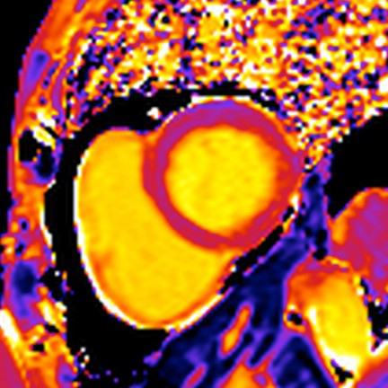
Introduction: Amyloidosis with cardiac involvement is often underdiagnosed and has a high mortality rate. Cardiac magnetic resonance imaging (MRI) with native T1 Mapping (without IV contrast) could serve as an early marker of the disease, but its diagnostic accuracy in our setting is unknown. Objectives: To determine the diagnostic value of native T1 Mapping in patients with cardiac light chain (AL) amyloidosis. Methods: Evaluation of Cardiac MRI as a diagnostc test in 8 patients with light chain amyloidosis (AL) with cardiac involvement and 18 healthy individuals.
All patients underwent 3T (Tesla) CMR with native T1 Mapping sequence (MOLLI, Modified Look- Locker Inversion-recovery) and longitudinal and short-axis cine images. The T1 mapping mean with 95 % CI was estimated and sensitivity and specificity were calculated. Inter and intra-observer agreement was evaluated. Results: Patients with AL amyloidosis demonstrated increased values of Native T1 Mapping compared to healthy ones (1,415 ms ± 69 vs. 1,214 ms ± 44; p <0.001). The value of the native T1 Mapping in patients with AL amyloidosis varied between 1,322 and 1,493 ms while in the healthy it varied between 1,098 and 1,285 ms. A native T1 Mapping value greater than 1,357 ms (3.25 standard deviations) achieves a sensitivity (S) of 87.5 %, a specificity (E) of 100 %, and an accuracy in the classification of sick or healthy of 96.2 %. Conclusions: The native T1 Mapping CMR has adequate diagnostic performance and is a sensitive and specific tool for identifying patients with AL cardiac amyloidosis.
Downloads
References
Wechalekar AD, Gillmore JD, Hawkins PN. Systemic amyloidosis. Lancet. 2016;387:2641-54.
Sharma N, Howlett J. Current state of cardiac amyloidosis. Curr Opin Cardiol. 2013;28:242-8.
Lousada I, Comenzo RL, Landau H. Light chain amyloidosis: patient experience survey from the Amyloidosis Research Consortium. Adv. Ther. 2015;32:920-8.
Grogan M, Dispenzieri , Gertz MA. Light-chain cardiac amyloidosis: strategies to promote early diagnosis and cardiac response. Heart. 2017;103:1065-72.
Falk RH, Alexander KM, Liao R. AL (Light-Chain) cardiac amyloidosis a review of diagnosis and therapy. JACC. 2016;68(12):1323-41.
Rubin J, Maurer MS. Cardiac amyloidosis: overlooked, underappreciated, and treatable. Annu. Rev. Med. 2020;71:203-19.
Sperry RW, Reyes BA, Ikram A, et al. Tenosynovial and cardiac amyloidosis in patients undergoing carpal tunnel release. J Am Coll Cardiol. 2018;72:2040-50.
Duque M, Velásquez JE, Marín JE. Amiloidosis cardiaca. Rev Colomb Cardiol. 2009;16:118-27.
Fontana M, Banypersad SM, Treibel T, Moon JC. Native T1 Mapping in transthyretin amyloidosis. JACC. 2014;7(2):157-65.
Everett RJ, Stirrat CG, Semple S. Assessment of myocardial fibrosis with T1 mapping MRI. Clin Radiol. 2016;71:768e778.
From AM, Maleszewski JJ. Current status of endomyocardial biopsy. Mayo Clin Proc. 2011;86(11):1095-102.
Ruberg FL. T1 mapping in cardiac amyloidosis can we get there from here? JACC. 2013;6(4):498-500.
Fontana N, Chung R, Hawkins PN, Moon JC. Cardiovascular magnetic resonance for amyloidosis. Heart Fail Rev. 2015;20:133-44.
Karamitsos TD, Piechnik SK, Moon JC. Noncontrast T1 mapping for the diagnosis of cardiac amyloidosis. JACC. 2013;6(4):4887.
Bulluck H, Maestrini V, Rosmini S, Moon JC. Myocardial T1 Mapping. Hope or hype? Circ J. 2015;79:487-94.
Lin L, Li X, Feng J, Shen K. The prognostic value of T1 mapping and late gadolinium enhancement cardiovascular magnetic resonance imaging in patients with light chain amyloidosis. J Cardiovasc Magn Reson. 2018;20(1):2.
Qian C, Jing Y, Wen S. Late gadolinium enhancement and T1 mapping for the diagnosis of cardiac amyloidosis. Chin Crit Care Med. 2019;31(12):1538-41.
Dabir D, Child N, Kalra A. Reference values for healthy human myocardium using a T1 mapping methodology: results from the International T1 Multicenter cardiovascular magnetic resonance study. J Cardiovasc Magn Reson. 2014;16(1):69.
Banypersad SM, Fontana M, Maestrini V. T1 mapping and survival in systemic lightchain amyloidosis. Eur Heart J. 2015;36:244-51.
Kramer CM, Chandrashekhar Y, Narula J. T1 Mapping by CMR in cardiomyopathy: A noninvasive myocardial biopsy. JACC. 2013;6(4):532-4.
Baggiano A, Boldrini M, Martinez-Naharro A. Noncontrast magnetic resonance for the diagnosis of cardiac amyloidosis. JACC. 2020;13(1 Pt 1):69-80.
Lin K, Suwa K, Ma H, Collins J. Variability of native T1 values: implication for defining regional myocardial changes using MRI. Int J Cardiovasc Imag. 2018;34(10):1637-45.
dem Siepen FA, Baumgärtner C, Müller-Henessen M, et al. Variability of cardiovascular magnetic resonance (CMR) T1 mapping parameters in healthy volunteers during long-term follow-up. Open Heart 2018;5(1):e000717.
Briasoulis A, Lama N, Rempakos A, et al. Diagnostic and prognostic value of nonlate gadolinium enhancement cardiac magnetic resonance parameters in cardiac amyloidosis. Curr Probl Cardiol. 2023;48(4):101573. https://doi.org/10.1016/j.cpcardiol.2022.101573.
Ioannou A, Patel RK, Martínez-Naharro A, et al. Tracking treatment response in cardiac light-chain amyloidosis with native T1 Mapping. JAMA. 2023;8(9):848-52. https://doi.org/10.1001/jamacardio.2023.2010.
Downloads
Published
How to Cite
Issue
Section
License
Copyright (c) 2026 Revista Colombiana de Radiología

This work is licensed under a Creative Commons Attribution-NonCommercial-ShareAlike 4.0 International License.
La Revista Colombiana de Radiología es de acceso abierto y todos sus artículos se encuentran libre y completamente disponibles en línea para todo público sin costo alguno.
Los derechos patrimoniales de autor de los textos y de las imágenes del artículo como han sido transferidos pertenecen a la Asociación Colombiana de Radiología (ACR). Por tanto para su reproducción es necesario solicitar permisos y se debe hacer referencia al artículo de la Revista Colombiana de Radiología en las presentaciones o artículos nuevos donde se incluyan.
| Article metrics | |
|---|---|
| Abstract views | 575 |
| Galley views | 196 |
| PDF Views | 196 |
| HTML views | 0 |
| Other views | 0 |