3D CMR–guided ventricular tachycardia ablation
DOI:
https://doi.org/10.53903/01212095.463Keywords:
Ventricular Tachycardia, Cardiac Magnetic Resonance Imaging, Catheter Ablation, Myocardial Fibrosis, ElectrophysiologyAbstract
Sustained monomorphic ventricular tachycardia (VT) is a potentially life-threatening arrhythmia, particularly prevalent in patients with structural heart disease and myocardial scarring, where reentry mechanisms predominate. Catheter ablation is a key therapeutic strategy; however, its effectiveness depends on accurate identification of the arrhythmogenic substrate. Cardiac magnetic resonance imaging (CMRI) with three-dimensional delayed enhancement (3D-DE) allows for a detailed anatomical characterization of myocardial fibrosis and borderzone channels. We present the case of a 52-year-old man with prior myocarditis, hypertension, and type 2 diabetes mellitus, carrying an implantable cardioverter-defibrillator, who was admitted due to multiple appropriate
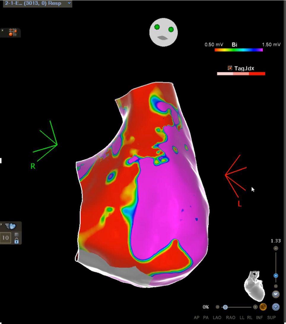
shocks. A 3T CMR study with 3D LGE and ADAS 3D analysis identified intramural septal fibrosis with biventricular border zone corridors. Electro-anatomical mapping confirmed low-voltage areas consistent with the three-dimensional reconstruction. Two VTs with different morphologies were induced, and their critical isthmuses were localized in right and left septal regions guided by CMRderived anatomical information. Targeted ablation resulted in termination of both arrhythmias, without complications or recurrences during follow-up. This case illustrates the clinical value of image–electrophysiology integration, highlighting how three-dimensional CMR substrate analysis may optimize ablation strategy in complex scar-related ventricular arrhythmias.
Downloads
References
Mehrotra AK, Callans D. Role of cardiac magnetic resonance imaging in the management and treatment of ventricular tachycardia in patients with structural heart disease. Circ J. 2015;79:1656-61. https://doi.org/10.1253/circj.CJ-15-0606
John RM, Tedrow UB, Koplan BA, Albert CM, Epstein LM, Sweeney MO, et al. Ventricular arrhythmias and sudden cardiac death. The Lancet. 2012;380:1520-9. https://doi.org/10.1016/S0140-6736(12)61413-5
Sapp JL, Tang ASL, Parkash R, Stevenson WG, Healey JS, Gula LJ, et al. Catheter ablation or antiarrhythmic drugs for ventricular tachycardia. N Engl J Med. 2025;392:737-47. https://doi.org/10.1056/NEJMoa2409501
Karamitsos TD, Francis JM, Myerson S, Selvanayagam JB, Neubauer S. The role of cardiovascular magnetic resonance imaging in heart failure. J Am Coll Cardiol. 2009;54:1407-24. https://doi.org/10.1016/j.jacc.2009.04.094
Zuluaga-Santamaría A, Aldana-Sepúlveda N, Duque-González L, Blanco-Daza E, Zuluaga-Molina N, Astudillo-Ramírez V, et al. Resonancia magnética cardiaca: utilidad de la secuencia de realce tardío 3D (RT-3D) en electrofisiología. Rev Colomb Cardiol.
2022;28:7806. https://doi.org/10.24875/RCCAR.M21000101
Muser D, Santangeli P, Selvanayagam JB, Nucifora G. Role of cardiac magnetic resonance imaging in patients with idiopathic ventricular arrhythmias. Curr Cardiol Rev. 2018;15:12-23. https://doi.org/10.2174/1573403X14666180925095923
Thomsen AF, Bertelsen L, Jøns C, Jabbari R, Lønborg J, Kyhl K, et al. Scar border zone mass and presence of border zone channels assessed with cardiac magnetic resonance imaging are associated with ventricular arrhythmia in patients with ST-segment elevation
myocardial infarction. EP Eur. 2023;25:978-88. https://doi.org/10.1093/europace/euac256
Pisani CF, Alexandre FK, Kulchetscki R, Mayrink M, Wu TC, Chokr M, et al. Initial experience on cardiac magnetic resonance-aided VT ablation in South America. J Interv Card Electrophysiol. 2023;66:1581-7. https://doi.org/10.1007/s10840-022-01464-x
Jang J, Hwang HJ, Tschabrunn CM, Whitaker J, Menze B, Anter E, et al. Cardiovascular magnetic resonance-based three-dimensional structural modeling and heterogeneous tissue channel detection in ventricular arrhythmia. Sci Rep. 2019;9:9317. https://doi.org/10.1038/s41598-019-45586-1
Amoni M, Vermoortele D, Ekhteraei-Tousi S, Doñate Puertas R, Gilbert G, Youness M, et al. Heterogeneity of repolarization and cell-cell variability of cardiomyocyte remodeling within the myocardial infarction border zone contribute to arrhythmia susceptibility. Circ Arrhythm Electrophysiol. 2023;16. https://doi.org/10.1161/CIRCEP.122.011677
Auricchio A, Faletra FF. Use of contemporary imaging techniques for electrophysiological and device implantation procedures. JACC Cardiovasc Imaging. 2020;13:851-65. https://doi.org/10.1016/j.jcmg.2019.01.043
Soto-Iglesias D, Penela D, Jáuregui B, Acosta J, Fernández-Armenta J, Linhart M, et al. Cardiac magnetic resonance-guided ventricular tachycardia substrate ablation. JACC Clin Electrophysiol. 2020;6:436-47. https://doi.org/10.1016/j.jacep.2019.11.004
Vázquez-Calvo S, Casanovas JM, Garre P, Ferró E, Sánchez-Somonte P, Quinto L, et al. Evolution of deceleration zones during ventricular tachycardia ablation and relation with cardiac magnetic resonance. JACC Clin Electrophysiol. 2023;9:779-89.
https://doi.org/10.1016/j.jacep.2022.12.015
Aoki H, Sato H, Fukuda-Shimada R, Kimura Y, Taguchi I, Nakahara S. Assessment of the ventricular tachycardia isthmus depth using 3-dimensional cardiac magnetic resonance imaging and peak frequency mapping in ischemic cardiomyopathy. Hear Case Rep. 2025;S2214027125003434. https://doi.org/10.1016/j.hrcr.2025.10.033
Arya A, Di Biase L, Bazán V, Berruezo A, d’Avila A, Della Bella P, et al. Epicardial ventricular arrhythmia ablation: a clinical consensus statement of the European Heart Rhythm Association of the European Society of Cardiology and the Heart Rhythm Society, the Asian Pacific Heart Rhythm Society, the Latin American Heart Rhythm Society, and the Canadian Heart Rhythm Society. Eur Eur Pacing Arrhythm Card Electrophysiol J Work Groups Card Pacing Arrhythm Card Cell Electrophysiol Eur Soc Cardiol. 2025;27:euaf055. https://doi.org/10.1093/europace/euaf055
Shamloo AS, Di Biase L, Arya A. Clinical consensus statement on epicardial ventricular arrhythmia ablation: key messages. Eur Heart J. 2026;47:148-9. https://doi.org/10.1093/eurheartj/ehaf741
Chery G, Khoshknab M, Nazarian S. Imaging to facilitate ventricular tachycardia ablation. JACC Clin Electrophysiol. 2024;10:2277-92. https://doi.org/10.1016/j.jacep.2024.08.003
Downloads
Published
How to Cite
Issue
Section
License
Copyright (c) 2026 Revista Colombiana de Radiología

This work is licensed under a Creative Commons Attribution-NonCommercial-ShareAlike 4.0 International License.
La Revista Colombiana de Radiología es de acceso abierto y todos sus artículos se encuentran libre y completamente disponibles en línea para todo público sin costo alguno.
Los derechos patrimoniales de autor de los textos y de las imágenes del artículo como han sido transferidos pertenecen a la Asociación Colombiana de Radiología (ACR). Por tanto para su reproducción es necesario solicitar permisos y se debe hacer referencia al artículo de la Revista Colombiana de Radiología en las presentaciones o artículos nuevos donde se incluyan.
| Article metrics | |
|---|---|
| Abstract views | 384 |
| Galley views | 185 |
| PDF Views | 185 |
| HTML views | 0 |
| Other views | 0 |