
Cortical Laminar Necrosis in Computed Tomography Scan: A Case Report
DOI:
https://doi.org/10.53903/01212095.97Keywords:
Cerebral cortex, Tomography, X-ray computed, Neuroimaging, Brain hypoxiaAbstract
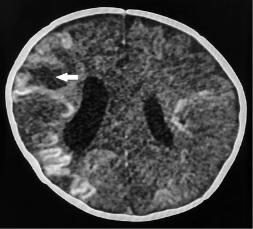
Introduction: Cortical laminar necrosis is an imaging term used to describe gyriform hyperdense lesions in the cortical region that can be appreciated on the CT (Computed Tomography). The etiology of this finding involves brain energy depletion, which can be derived from multiple pathologies, that mainly lead to hypoxia or metabolic alterations. Case report: the case of a 3-month-old female patient is presented; she was taken to the emergency department with a 1-week clinical manifestations consisting of thermal rises, dyspnea, cyanosis coldness in extremities, tachypnea, tachycardia and edema. An echocardiography diagnosed myocarditis of probable viral origin. Subsequently, the patient presented cardiac arrest and prolonged shock that required cardiopulmonary resuscitation and admission to neonatal ICU. A cerebral CT scan taken 1 week after these events evidenced imaging findings compatible with cortical laminar necrosis. Discussion: There are no epidemiological descriptions of this finding, however, an increase has been theorized due to the high survival rates of patients suffering from ischemic hypoxic pathologies. Although findings of cortical laminar necrosis are typical of MRI, they can also be seen on CT scan, where they typically show as a subtle cortical hyperdensity, and the most striking findings are associated to a worst prognosis. Conclusion: Disclosure of these type of radiological images is necessary in order to promote more accurate prognoses in patients in whom these findings are appreciated.
Downloads
References
Niwa T, Aida N, Shishikura A, et al. Susceptibility-weighted imaging findings of cortical laminar necrosis in pediatric patients. Am J Neuroradiol. 2008;9:1795-8.
Garg RK, Rizvi I, Ingole R, et al. Cortical laminar necrosis in dengue encephalitis—a case report. BMC Neurology. 2017;17(1).
De la Riva P, Maneiro M, Martí-Massó J, et al. Encefalopatía hipóxico-isquémica: lesiones en resonancia magnética. Neurología. 2011;26(6):371-2.
Samain JL, Haven F, Gille M, et al. Typical CT and MRI features of cortical laminar necrosis. J Belgian Soc Radiol. 2011;94(6):357.
Renard D, Castelnovo G, Bouly S, et al. Cortical abnormalities on MRI: what a neurologist should know. Pract Neurol. 2015;15:257-65.
Tibussek D, DeVeber G, Yau I, et al. PP04.15-2395: Cortical laminar necrosis after neonatal and childhood arterial ischemic stroke. A relevant finding? Eur J Paediatric Neurol. 2015;19(Supplement 1):S45.
Serrano M, Ara JR, Fayed M, et al. Encefalopatía hipóxica y necrosis laminar cortical. Rev Neurol. 2001;32(9):843-7.
Sawada H, Udaka F, Seriu N, et al. MRI demonstration of cortical laminar necrosis and delayed white matter injury in anoxic encephalopathy. Neuroradiology. 1990;32(4):319-21.
Downloads
Published
How to Cite
Issue
Section
License

This work is licensed under a Creative Commons Attribution-NonCommercial-ShareAlike 4.0 International License.
La Revista Colombiana de Radiología es de acceso abierto y todos sus artículos se encuentran libre y completamente disponibles en línea para todo público sin costo alguno.
Los derechos patrimoniales de autor de los textos y de las imágenes del artículo como han sido transferidos pertenecen a la Asociación Colombiana de Radiología (ACR). Por tanto para su reproducción es necesario solicitar permisos y se debe hacer referencia al artículo de la Revista Colombiana de Radiología en las presentaciones o artículos nuevos donde se incluyan.


