Masas sólidas y quísticas inguinales en mujeres. Presentación de caso y revisión de tema
DOI:
https://doi.org/10.53903/01212095.349Palabras clave:
Quiste epidermoide, Región inguinal, Canal inguinalResumen
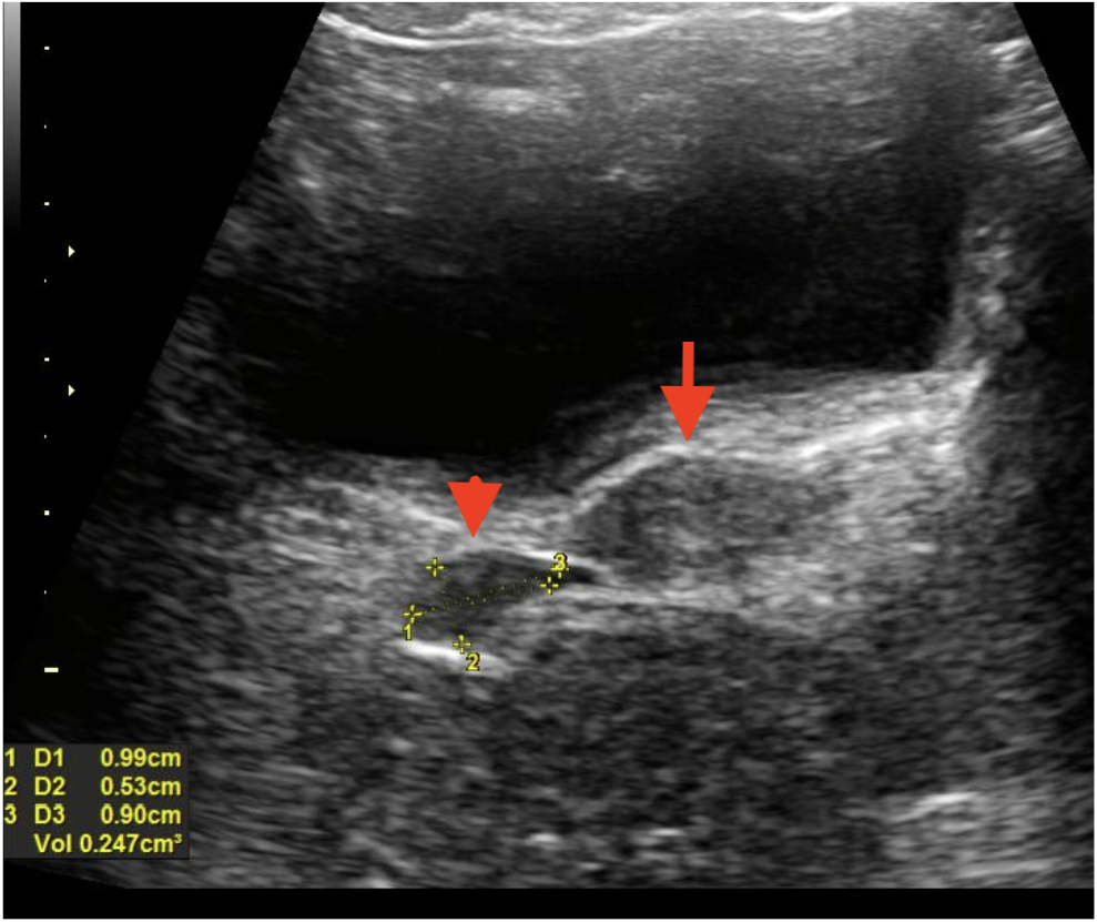
Las masas inguinales involucran un número importante de diagnósticos diferenciales. Tienen manifestaciones clínicas distintas, particularmente malestar y angustia psicosocial. Es crucial conocer la anatomía, embriología y fisiología asociadas con la formación de anomalías, especialmente en mujeres, para comprender su inicio y progresión, entre estas, de las alteraciones asociadas al canal de Nuck, el cual es la contraparte femenina del proceso peritoneo-vaginal en el hombre. Como parte del desarrollo normal, su obliteración se da durante el primer año de vida; sin embargo, hay ocasiones en las que, de manera anómala, se producen hidroceles comunicantes o quistes en su extensión que se manifiestan como masas inguinales. Sin embargo, no se deben olvidar los diagnósticos diferenciales. Se hizo una búsqueda de la literatura en PubMed utilizando palabras clave para obtener la información relevante. Se presenta un caso clínico y las características principales de los diagnósticos diferenciales.
Descargas
Referencias bibliográficas
Chihara N, Taniai N, Suzuki H, Nakata R, Shioda M, Yoshida H. Use of a novel open posterior wall technique for laparoscopic excision of hydrocele of the canal of Nuck in an adult female: case report. J Nippon Med School. 2019;86(6):345-8. http://dx.doi.org/10.1272/jnms.JNMS.2019_86-601
Topal U, Sarıtaş AG, Ülkü A, Akçam AT, Doran F. Cyst of the canal of Nuck mimicking inguinal hernia. Int J Surg Case Rep [Internet]. 2018;52:117-9. http://dx.doi.org/10.1016/j.ijscr.2018.09.053
Yen CF, Wang CJ, Lin SL, Chang PC, Lee CL, Soong YK. Laparoscopic closure of patent canal of Nuck for female indirect inguinal hernia. J Am Assoc Gynecol Laparosc [Internet]. 2001;8(1):143-6. http://dx.doi.org/10.1016/s1074-3804(05)60564-6
Counseller VS, Black BM. Hydrocele of the canal of nuck: Report of seventeen cases. Ann Surg [Internet]. 1941;113(4):625-30. http://dx.doi.org/10.1097/00000658-194104000-00012
Wang L, Maejima T, Fukahori S, Shun K, Yoshikawa D, Kono T. Laparoscopic surgical treatment for hydrocele of canal of Nuck: A case report and literature review. Surg Case Rep [Internet]. 2021;7(1):121. http://dx.doi.org/10.1186/s40792-021-01205-8
Cheng EM, Sarkar A, Perera DS. Hydrocoele in the canal of Nuck in an adult female: a rare cause for inguinal swelling. ANZ J Surg [Internet]. 2021;91(4):E210-1. http://dx.doi.org/10.1111/ans.16279
Kaeser MA, Haun DW, Cho JCS, Kettner NW. Hydrocele in the Canal of Nuck. J Med Ultrasound [Internet]. 2011;19(4):138-40. http://dx.doi.org/10.1016/j.jmu.2011.10.001
Kim BM, Lee JY, Han YH, Kim SY, Seo JW, Kim YH, et al. Mesothelial cyst of the round ligament mimicking a metastasis: a case report. Korean J Radiol [Internet].2010;11(3):364-7. http://dx.doi.org/10.3348/kjr.2010.11.3.364
Vargas-Ávila AL, Gómez-Montoya LE, Guidos-Gil JC, Ávila-Rivera JL, Huerta-García de León Ó, Medina-Tirado MA. Round ligament cyst simulating incarcerated inguinal hernia. Report of a case. Cir Cir [Internet]. 2017;85(4):334-8. http://dx.doi.
org/10.1016/j.circir.2016.03.008
Mine Y, Eguchi S, Enjouji A, Fukuda M, Yamaguchi J, Inoue Y, et al. Round ligament varicosities diagnosed as inguinal hernia during pregnancy: A case report and series from two regional hospitals in Japan. Int J Surg Case Rep [Internet]. 2017;36:122-5. http://dx.doi.org/10.1016/j.ijscr.2017.05.006
Oh SN, Jung SE, Rha SE, Lim GY, Ku YM, Byun JY, et al. Sonography of various cystic masses of the female groin. J Ultrasound Med [Internet]. 2007;26(12):1735-42. http://dx.doi.org/10.7863/jum.2007.26.12.1735
Hertzberg BS, Middleton WD. Chapter 9: General abdomen. En: Hertzberg BS, Middleton WD, editores. Ultrasound: The Requisites. 3ª ed. Filadelfia, PA, Estados Unidos de América: Elsevier - Health Sciences Division; 2015. p. 204-28.
Tatarchuk T, Kosei N, Bachynska I, Yevtushenko N, Glamazda M. 129 uterine leiomyoma in a 16-year-old girl: Case report. Eur J Obstet Gynecol Reprod Biol [Internet]. 2022;273:e54-5. http://dx.doi.org/10.1016/j.ejogrb.2022.02.158
Bennett GL. Evaluación del dolor pélvico en las pacientes en edad fértil. En: Norton ME, Callen PW, editores. Ecografía en obstetricia y ginecología. Elsevier Health Sciences; 2017 Nov 24. p. 883-918.
Kamaya A, Wong-You-Cheong J, Woodward PJ, Kennedy AM, Sohaey R, Yuan X, et al. Bartholin cyst. En: Kamaya A, Wong-You-Cheong J, Woodward PJ, Kennedy AM, Sohaey R, Yuan X, Del Barco OR, Burke J, editores. Diagnostic Ultrasound for Sonographers E-book. Elsevier; 2019. p. 345.
Wortsman X. Técnicas de diagnóstico por imagen ecográfica en dermatología. En: Hamblin MR, Avci P, Gupta GK, editores. Diagnóstico por imagen en dermatología. Elsevier; 2018. p. 357-74.
Beverly S, Bronner J, Tayal V. Ultrasonographic detection of a high grade sarcoma in the emergency department. Vis J Emerg Med [Internet]. 2016;3:19-21. http://dx.doi.org/10.1016/j.visj.2015.11.007
Zito PM, Scharf R. Epidermoid cyst. En: StatPearls. Treasure Island (FL): StatPearls Publishing; 2025. https://www.ncbi.nlm.nih.gov/books/NBK499974/
Descriptores en Ciencias de la Salud: DeCS [Internet]. ed. 2025. Sao Paulo (SP): BIREME / OPS / OMS. 2025 [actualizado 2025 mar 28; citado 2025 agosto 30). Disponible en: https://decs.bvsalud.org/es/ths/resource/?id=4895&filter=ths_termall&q=quiste%20epidermoide#Details
Uzun I, İnan C, Varol F, Erzincan S, Sütcü H, Sayin C. Hemorrhagic cyst of the canal of Nuck after vaginal delivery presenting as a painful inguinal mass in the early postpartum period. Eur J Obstet Gynecol Reprod Biol [Internet]. 2017;213:147-8. http://dx.doi.org/10.1016/j.ejogrb.2017.04.020
Stickel WH, Manner M. Female hydrocele (cyst of the canal of nuck): Sonographic appearance of a rare and little-known disorder. J Ultrasound Med [Internet]. 2004;23(3):429-32. http://dx.doi.org/10.7863/jum.2004.23.3.429
Nasser H, King M, Rosenberg HK, Rosen A, Wilck E, Simpson WL. Anatomy and pathology of the canal of Nuck. Clin Imaging [Internet]. 2018;51:83-92. http://dx.doi.org/10.1016/j.clinimag.2018.02.003
Patel B, Zivin S, Panchal N, Wilbur A, Bresler M. Sonography of female genital hernias presenting as labia majora masses. J Ultrasound Med [Internet]. 2014;33(1):155-9. http://dx.doi.org/10.7863/ultra.33.1.155
Ferreira AF, Marques JP, Falcão F. Hydrocele of the canal of Nuck presenting as a sausage-shaped mass. Case Reports. 2017;2017:bcr-2017. http://dx.doi.org/10.1136/bcr-2017-221024
Descargas
Publicado
Cómo citar
Número
Sección
Licencia
Derechos de autor 2026 Revista colombiana de radiología

Esta obra está bajo una licencia internacional Creative Commons Atribución-NoComercial-CompartirIgual 4.0.
La Revista Colombiana de Radiología es de acceso abierto y todos sus artículos se encuentran libre y completamente disponibles en línea para todo público sin costo alguno.
Los derechos patrimoniales de autor de los textos y de las imágenes del artículo como han sido transferidos pertenecen a la Asociación Colombiana de Radiología (ACR). Por tanto para su reproducción es necesario solicitar permisos y se debe hacer referencia al artículo de la Revista Colombiana de Radiología en las presentaciones o artículos nuevos donde se incluyan.
| Estadísticas de artículo | |
|---|---|
| Vistas de resúmenes | 471 |
| Vistas de PDF | 583 |
| Descargas de PDF | 583 |
| Vistas de HTML | 0 |
| Otras vistas | 0 |