Signos sugestivos de lesiones osteocondrales en resonancias de rodilla
DOI:
https://doi.org/10.53903/01212095.401Palabras clave:
Imagen por resonancia magnetica, Rodilla, Cartílago articular, OsteocondritisResumen
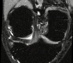
Introducción: La lesión osteocondral es una anormalidad del cartílago articular y del hueso subcondral, que puede ser causada por una variedad de entidades agudas y crónicas. Dichas lesiones se manifiestan en resonancia magnética desde discretos cambios de intensidad de señal del cartílago articular hasta pérdida de la morfología asociada a edema óseo. Objetivos: Determinar la frecuencia y caracterizar cuantitativamente (mediante el volumen del edema y el área del defecto condral) las lesiones osteocondrales en pacientes sintomáticos evaluados con RM de rodilla; describir la localización más frecuente de estas lesiones y evaluar la asociación estadística con patologías concomitantes como las lesiones meniscales y del ligamento cruzado anterior. Metodología: Se recolectaron las características epidemiológicas y radiológicas en los pacientes que acudieron a realizarse RM de rodilla en una clínica privada en Caracas en el 2022. Se identificaron signos sugestivos de lesiones osteocondrales en 477 estudios. Se clasificó el volumen del edema óseo subcondral dividiendo a los pacientes en 3 grupos: leve (0-3.000 mm3), moderado (3.001-10.000 mm3) y grave (>10.000 mm3). Resultados y conclusiones: De 477 estudios de rodilla, 229 (48 %) demostraron edema subcondral. El grupo etario con mayor frecuencia de edema subcondral osciló entre 41-60 años. La rótula resultó ser el lugar más frecuente con edema subcondral leve; sin embargo, los edemas moderados y graves se localizaron con mayor frecuencia en el cóndilo interno (20,7 %) y en el platillo tibial interno (19 %). La afectación cartilaginosa estuvo presente en un 82,5 % de los estudios positivos. El diagnóstico precoz de las lesiones osteocondrales puede ayudar al médico tratante a disminuir su progresión o incluso revertir el edema óseo antes de que la lesión progrese a un defecto cartilaginoso grave.
Descargas
Referencias bibliográficas
Chien A, Weaver J, Kinne E, Omar I. Magnetic resonance imaging of the knee. Pol J Radiol. 2020;85:e509-e531. https://doi.org/10.5114/pjr.2020.99415.
Trattnig S, Millington A, Szomolanyi P, Marlovits S. MR imaging of osteochondral grafts and autologous chondrocyte implantation. Eur Radiol. 2007;17(1):103-18. https://doi.org/10.1007/s00330-006-0333-z.
Gorbachova T, Melenevsky Y, Cohen M, Cerniglia B. Osteochondral lesions of the knee: differentiating the most common entities at MRI. Radiographics. 2018;38(5):1478-95. https://doi.org/10.1148/rg.2018180044.
Huizinga J, Shah N, Smith S, Notino A, Kluczynski M. Prevalence of undiagnosed subchondral insufficiency fractures of the knee in middle age adults with knee pain and suspected meniscal tear. Osteoarthr Cartil Open. 2020;2(4):100089. https://doi.org/10.1016/j.ocarto.2020.100089.
Zanetti M, Bruder E, Romero J, Hodler J. Bone marrow edema pattern in osteoarthritic knees: correlation between MR imaging and histologic findings. Radiology. 2000;215(3):835-40. https://doi.org/10.1148/radiology.215.3.r00jn05835.
Vallejo M, Román P, Gómez-Millán P. Rodilla: indicaciones, protocolo de estudio, anatomia normal y variantes anatómicas. 2nd ed. Panamericana; 2023.
Fröhlich S, Peterhans L, Stern C, Frey W, Sutter R, Spörri J. Remarkably high prevalence of overuse related knee complaints and MRI abnormalities in youth competitive alpine skiers: a descriptive investigation in 108 athletes aged 13–15 years. BMJ Open Sp Ex Med. 2020;6(1):e000738. https://doi.org/10.1136/bmjsem-2020-000738.
Geith T, Stellwag A, Müller P, Reiser M, Baur-Melnyk A. Is bone marrow edema syndrome a precursor of hip or knee osteonecrosis? Results of 49 patients and review of the literature. Diagn Interv Radiol. 2020(4):355-62. https://doi.org/10.5152/dir.2020.19188.
García-Valtuille R, Abascal F, Sampedro J, Pesquera L. RM de la rodilla. Atlas de los principales halazgos por RM en la patología de la rodilla. OSATEK, SA. https://dx.doi.org/10.1594/seram2014/S-0943
Marinetti A, Tessarolo F, Ventura L. Morphological MRI of kneww cartilage: repeatability and reproducibility of damage evaluation and correlation with gross pathology examination. Eur Radiol. 2020(6):3226-35. https://doi.org/10.1007/s00330-019-06627-5.
ICRS. International Cartilage Regeneration & Joint Preservation Society [internet]. [citado: 2025 jul 02]. Disponible en: https://cartilage.org/icrs-score-grade/.
Aroen A, Loken S, Heir S, Alvik E, Ekeland A. Articular cartilage lesions in 993 consecutive knee arthroscopies. Am J Sports Med. 2004;32(1):211-5. https://doi.org/10.1177/0363546503259345.
Streitparth F, Schottle P, Schlichting K, Fischbach F, Denecke T. Osteochondral defect repair after implantation of biodegradable scaffolds: indirect magnetic resonance arthrography and histopatologic correlation. Acta Radiol. 2009;50(7):765-74. https://doi.org/10.1080/02841850902980272.
Mini NP, Chung CB, Resnick DL. Acute and Stress-related injuries of bones and cartilage: pertinent anatomy, basic biomechanics, and imaging perspective. Radiology. 2016;280(1):21-38. https://doi.org/10.1148/radiol.16142305.
Zicaro JP, Molina A, Yacuzzi C, Costa M. Trasplante osteocondral autólogo para tratar lesiones osteocondrales de la rodilla: evaluación de 62 pacientes con un seguimiento promedio de ocho años. Rev. Asoc. Argent. Ortop. Traumatol. [internet]. 2018 Jun [citado: 2026 feb 22];83(2):107-12. Disponible en: https://www.scielo.org.ar/scielo.php?script=sci_arttext&pid=S1852-74342018000200007&lng=es.
Gaillard F, Knipe H, Jones J. Radiopaedia.org. [Online]. Available from: https://radiopaedia.org/articles/1120
Gaillard F, Jones J, Botz B. Radiopaedia.org. [Online]. Available from: https://radiopaedia.org/articles/1796
Álvarez A, García L, García Y, Gutiérrez M. Osteocondritis disecante del astrágalo. Revisión del tema. 2003;7:646-56.
Helms C, Major N, Anderson M, Kaplan P, Dussault R. RM musculoesquelética. MARBAN; 2000.
Álvarez E, Ripoll P, Restrepo A, Forriol F. Revisión de la reparación del cartílago. Posibilidades y resultados. Trauma Fund MAPFRE. 2010;21(2):117-34.
Tas M, Avinca Ö, Şen A, Ari B, Tuzun C. Magnetic resonance imaging (MRI) findings after knee, foot, and ankle traumas. Eur Rev Med Pharmacol Sci. 2022;26(23):8675-83. https://doi.org/10.26355/eurrev_202212_30537.
Mayerhoefer M, Breitenseher M, Hofmann S, Aigner N, Meizer R. Computer-assisted quantitative analysis of bone marrow edema of the knee: initial experience with a new method. AJR. 2004;182(6):1399-403. https://doi.org/10.2214/ajr.182.6.1821399.
Tanamas S, Wluka A, Pelletier J, Abram F. Bone marrow lesions in people with knee osteoarthritis predict progression of disease and joint replacement: a longitudinal study. Rheumatology (Oxford). 2010;49(12):2413-9. https://doi.org/10.1093/rheumatology/keq286.
Moradi K, Mohammadi S, Roemer F, Momtazmanesh S, Hathaway Q. Progression of bone marrow lesions and the development of knee osteoarthritis: osteoarthritis initiative data. Radiology. 2024;312(3):e240470. https://doi.org/10.1148/radiol.240470.
Felson D, Chaisson C, Hill C, Totterman S, Gale M. The association of bone marrow lesions with pain in knee osteoarthritis. Ann Intern Med. 2001;134(7):541-9. https://doi.org/10.7326/0003-4819-134-7-200104030-00007.
Curl W, Krome J, Gordon S, Rushing J, Paterson B, Poehling G. Cartilage injuries: a review of 31,516 knee arthroscopies. Arthroscopic. 1997;13(4):456-60. https://doi.org/10.1016/s0749-8063(97)90124-9.
Menetrey J, Unno-Veith F, Madry H, Van Breuseghem I. Epidemiology and imaging of the subchondral bone in articular cartilage repair. Knee Sug Sports Traumatol Arthrosc. 2010;18(4):463-71. https://doi.org/10.1007/s00167-010-1053-0.
Zamber Rw, Teitz CC, McGuire DA, Frost JD, Hermanson BK. Articular cartilage lesions of the knee. Arthroscopy. 1989;5(4):258-68. https://doi.org/10.1016/0749-8063(89)90139-4.
Culvenor A, Elin B, Hart H, Stefanik J, Guermazi A, Crossley K. Prevalence of knee osteoarthritis features on magnetic resonance imaging in asymptomatic uninjured adults a systematic review and meta analysis. Br J Sports Med. 2019;53:1268-78.
https://doi.org/10.1136/bjsports-2018-099257.
Hjelle K, Solheim E, Strand T, Muri R, Brittberg M. Articular cartilage defects in 1,000 knee arthroscopies. Arthroscopy. 2002;18(7):730-4. https://doi.org/10.1053/jars.2002.32839.
Descargas
Publicado
Cómo citar
Número
Sección
Licencia
Derechos de autor 2026 Revista colombiana de radiología

Esta obra está bajo una licencia internacional Creative Commons Atribución-NoComercial-CompartirIgual 4.0.
La Revista Colombiana de Radiología es de acceso abierto y todos sus artículos se encuentran libre y completamente disponibles en línea para todo público sin costo alguno.
Los derechos patrimoniales de autor de los textos y de las imágenes del artículo como han sido transferidos pertenecen a la Asociación Colombiana de Radiología (ACR). Por tanto para su reproducción es necesario solicitar permisos y se debe hacer referencia al artículo de la Revista Colombiana de Radiología en las presentaciones o artículos nuevos donde se incluyan.
| Estadísticas de artículo | |
|---|---|
| Vistas de resúmenes | 1077 |
| Vistas de PDF | 332 |
| Descargas de PDF | 332 |
| Vistas de HTML | 0 |
| Otras vistas | 0 |