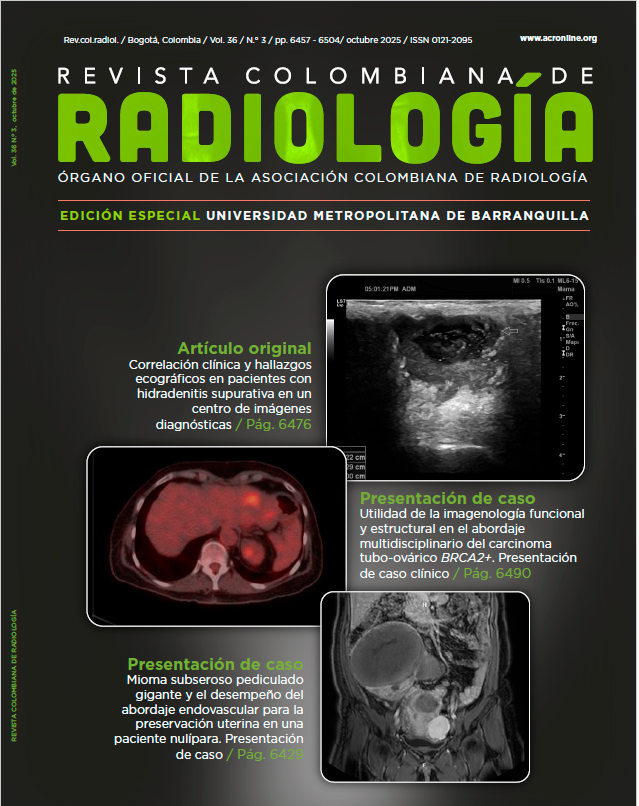
Utility of functional and structural imaging in the multidisciplinary management of BRCA2-positive tubo-ovarian carcinoma. A clinical case report
DOI:
https://doi.org/10.53903/01212095.398Keywords:
BRCA2 genes, Ovarian neoplasms, Positron-emission tomography computed tomography, Magnetic resonance imagingAbstract
Introduction: Carcinoma of the fallopian tube linked to BRCA2 mutations presents a significant clinical challenge that has transformed its management through the implementation of targeted therapies and genomic-based precision medicine. In this framework, both functional and structural imaging are essential, serving not only to aid in staging and monitoring treatment efficacy, but also to critically assess atypical findings that could impact therapeutic decision-making. Case presentation: A 63-year-old woman with a history of metachronous bilateral breast cancer was diagnosed with high-grade serous tubo-ovarian carcinoma with a germline BRCA2 mutation. She received platinum-based neoadjuvant chemotherapy, underwent complete cytoreductive surgery, and was initiated on maintenance therapy with Olaparib. During follow-up, a PET-CT revealed a focal uptake in hepatic segment II, initially suggestive of recurrence. Further assessment
with hepatobiliary contrast-enhanced MRI and image-guided liver biopsy ruled out malignancy. The patient remains on active surveillance with no evidence of disease recurrence. Discussion: This case highlights the clinical utility of integrating functional and structural
imaging in the personalized oncologic approach. Multimodal interpretation avoided unnecessary interventions and enabled the continuation of targeted therapy. The case emphasizes the value of multidisciplinary collaboration among oncology, radiology, and interventional imaging in validating suspicious findings that may otherwise lead to overtreatment. Conclusion: The correlation between PET-CT, liver MRI, and image-guided biopsy was essential in excluding tumor progression, underscoring the central role of radiology in clinical decision-making. This multidisciplinary strategy ensured optimal therapeutic continuity and reflects the impact of imaging in the era of precision oncology.
Downloads
References
Torre LA, Trabert B, DeSantis CE, et al. Ovarian cancer statistics, 2018. CA Cancer J Clin. 2018;68(4):284-96.
Moore K, Colombo N, Scambia G, et al. Maintenance Olaparib in patients with newly diagnosed advanced ovarian cancer. N Engl J Med. 2018;379(26):2495-505.
Ray-Coquard I, Pautier P, Pignata S, et al. Olaparib plus bevacizumab as maintenance therapy in ovarian cancer. N Engl J Med. 2019;381(25):2416-28.
Antoch G, Saoudi N, Kuehl H, et al. Accuracy of whole-body dual-modality fluorine- 18–2-fluoro-2-deoxy-D-glucose positron emission tomography and computed tomography (FDG-PET/CT) for tumor staging in solid tumors. J Clin Oncol. 2004;22(21):4357-68.
Metser U, Even-Sapir E. Increased (18)F-FDG uptake in benign, nonphysiologic lesions found on whole-body PET/CT: accumulated data from four years of experience with PET/CT. Semin Nucl Med. 2007;37(3):206-22.
Vogl TJ, Klinger C, Schwarz W, et al. Liver metastases: comparison of MR imaging with Gd-EOB-DTPA and Gd-DTPA. Radiology. 2011;258(3):766-76.
Seale MK, Catalano OA, Saini S, et al. Hepatobiliary-specific MR contrast agents: role in imaging the liver and biliary tree. Radiographics. 2009;29(6):1725-48.
Banerjee S, Gonzalez-Martin A, Harter P, et al. First-line PARP inhibitors in ovarian cancer: summary of evidence and future directions. Nat Rev Clin Oncol. 2020;17(11):666-81.
Park SH, Moon MH, Kim HJ, et al. Role of follow-up PET/CT in patients with recurrent ovarian cancer. Gynecol Oncol. 2008;109(2):274-80.
Sironi S, Bagnoli P, Garlaschi A, et al. Pelvic MRI with diffusion-weighted imaging in patients with suspected ovarian cancer: diagnostic value of ADC and correlation with histological grading. Eur Radiol. 2012;22(4):766-72.
Colombo N, Sessa C, du Bois A, et al. ESMO-ESGO consensus conference recommendations on ovarian cancer. Ann Oncol. 2019;30(5):672-705.
Hricak H, Gatsonis C, Chi DS, et al. Role of imaging in the management of ovarian cancer. Radiology. 2013;266(3):665-81.
Downloads
Published
How to Cite
Issue
Section
License
Copyright (c) 2026 Revista Colombiana de Radiología

This work is licensed under a Creative Commons Attribution-NonCommercial-ShareAlike 4.0 International License.
La Revista Colombiana de Radiología es de acceso abierto y todos sus artículos se encuentran libre y completamente disponibles en línea para todo público sin costo alguno.
Los derechos patrimoniales de autor de los textos y de las imágenes del artículo como han sido transferidos pertenecen a la Asociación Colombiana de Radiología (ACR). Por tanto para su reproducción es necesario solicitar permisos y se debe hacer referencia al artículo de la Revista Colombiana de Radiología en las presentaciones o artículos nuevos donde se incluyan.
| Article metrics | |
|---|---|
| Abstract views | 108 |
| Galley views | 45 |
| PDF Views | 45 |
| HTML views | 0 |
| Other views | 0 |